TED日本語
TED Talks(英語 日本語字幕付き動画)
TED日本語 - サンギータ・バティア: 体内で腫瘍を見つけ出すミクロの粒子
TED Talks
体内で腫瘍を見つけ出すミクロの粒子
This tiny particle could roam your body to find tumors
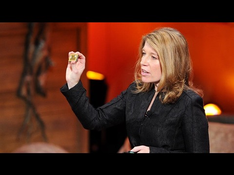
サンギータ・バティア
Sangeeta Bhatia
内容
もし、高価な検査施設や安定した電気の供給が無くても、悪性腫瘍を何年も前に見つけることができたらどうでしょう?医師、生体工学研究者、そして起業家の顔を持つサンギータ・バティアは、数々の学問領域を横断する研究室を率いて、人間の疾患を理解し、診断し、治療する革新的な方法を探しています。癌による死の3分の2は完全に予防可能であり、彼女の目標は、それを防ぐことだと言います。バティアが複雑なナノ粒子科学をずば抜けた明晰さで解説し、何百万もの命を救うであろう革新的な癌検診法について語ります。


字幕
SCRIPT
Script
In the space that used to house one transistor, we can now fit one billion. That made it so that a computer the size of an entire room now fits in your pocket. You might say the future is small.
As an engineer, I'm inspired by this miniaturization revolution in computers. As a physician, I wonder whether we could use it to reduce the number of lives lost due to one of the fastest-growing diseases on Earth: cancer. Now when I say that, what most people hear me say is that we're working on curing cancer. And we are. But it turns out that there's an incredible opportunity to save lives through the early detection and prevention of cancer.
Worldwide, over two-thirds of deaths due to cancer are fully preventable using methods that we already have in hand today. Things like vaccination, timely screening and of course, stopping smoking. But even with the best tools and technologies that we have today, some tumors can't be detected until 10 years after they've started growing, when they are 50 million cancer cells strong. What if we had better technologies to detect some of these more deadly cancers sooner, when they could be removed, when they were just getting started?
Let me tell you about how miniaturization might get us there. This is a microscope in a typical lab that a pathologist would use for looking at a tissue specimen, like a biopsy or a pap smear. This $ 7,000 microscope would be used by somebody with years of specialized training to spot cancer cells. This is an image from a colleague of mine at Rice University, Rebecca Richards-Kortum. What she and her team have done is miniaturize that whole microscope into this $ 10 part, and it fits on the end of an optical fiber. Now what that means is instead of taking a sample from a patient and sending it to the microscope, you can bring the microscope to the patient. And then, instead of requiring a specialist to look at the images, you can train the computer to score normal versus cancerous cells.
Now this is important, because what they found working in rural communities, is that even when they have a mobile screening van that can go out into the community and perform exams and collect samples and send them to the central hospital for analysis, that days later, women get a call with an abnormal test result and they're asked to come in. Fully half of them don't turn up because they can't afford the trip. With the integrated microscope and computer analysis, Rebecca and her colleagues have been able to create a van that has both a diagnostic setup and a treatment setup. And what that means is that they can do a diagnosis and perform therapy on the spot, so no one is lost to follow up.
That's just one example of how miniaturization can save lives. Now as engineers, we think of this as straight-up miniaturization. You took a big thing and you made it little. But what I told you before about computers was that they transformed our lives when they became small enough for us to take them everywhere. So what is the transformational equivalent like that in medicine? Well, what if you had a detector that was so small that it could circulate in your body, find the tumor all by itself and send a signal to the outside world? It sounds a little bit like science fiction. But actually, nanotechnology allows us to do just that. Nanotechnology allows us to shrink the parts that make up the detector from the width of a human hair, which is 100 microns, to a thousand times smaller, which is 100 nanometers. And that has profound implications.
It turns out that materials actually change their properties at the nanoscale. You take a common material like gold, and you grind it into dust, into gold nanoparticles, and it changes from looking gold to looking red. If you take a more exotic material like cadmium selenide -- forms a big, black crystal -- if you make nanocrystals out of this material and you put it in a liquid, and you shine light on it, they glow. And they glow blue, green, yellow, orange, red, depending only on their size. It's wild! Can you imagine an object like that in the macro world? It would be like all the denim jeans in your closet are all made of cotton, but they are different colors depending only on their size.
(Laughter)
So as a physician, what's just as interesting to me is that it's not just the color of materials that changes at the nanoscale; the way they travel in your body also changes. And this is the kind of observation that we're going to use to make a better cancer detector.
So let me show you what I mean. This is a blood vessel in the body. Surrounding the blood vessel is a tumor. We're going to inject nanoparticles into the blood vessel and watch how they travel from the bloodstream into the tumor. Now it turns out that the blood vessels of many tumors are leaky, and so nanoparticles can leak out from the bloodstream into the tumor. Whether they leak out depends on their size. So in this image, the smaller, hundred-nanometer, blue nanoparticles are leaking out, and the larger, 500-nanometer, red nanoparticles are stuck in the bloodstream. So that means as an engineer, depending on how big or small I make a material, I can change where it goes in your body.
In my lab, we recently made a cancer nanodetector that is so small that it could travel into the body and look for tumors. We designed it to listen for tumor invasion: the orchestra of chemical signals that tumors need to make to spread. For a tumor to break out of the tissue that it's born in, it has to make chemicals called enzymes to chew through the scaffolding of tissues. We designed these nanoparticles to be activated by these enzymes. One enzyme can activate a thousand of these chemical reactions in an hour. Now in engineering, we call that one-to-a-thousand ratio a form of amplification, and it makes something ultrasensitive. So we've made an ultrasensitive cancer detector.
OK, but how do I get this activated signal to the outside world, where I can act on it? For this, we're going to use one more piece of nanoscale biology, and that has to do with the kidney. The kidney is a filter. Its job is to filter out the blood and put waste into the urine. It turns out that what the kidney filters is also dependent on size. So in this image, what you can see is that everything smaller than five nanometers is going from the blood, through the kidney, into the urine, and everything else that's bigger is retained. OK, so if I make a 100-nanometer cancer detector, I inject it in the bloodstream, it can leak into the tumor where it's activated by tumor enzymes to release a small signal that is small enough to be filtered out of the kidney and put into the urine, I have a signal in the outside world that I can detect.
OK, but there's one more problem. This is a tiny little signal, so how do I detect it? Well, the signal is just a molecule. They're molecules that we designed as engineers. They're completely synthetic, and we can design them so they are compatible with our tool of choice. If we want to use a really sensitive, fancy instrument called a mass spectrometer, then we make a molecule with a unique mass. Or maybe we want make something that's more inexpensive and portable. Then we make molecules that we can trap on paper, like a pregnancy test. In fact, there's a whole world of paper tests that are becoming available in a field called paper diagnostics.
Alright, where are we going with this? What I'm going to tell you next, as a lifelong researcher, represents a dream of mine. I can't say that's it's a promise; it's a dream. But I think we all have to have dreams to keep us pushing forward, even -- and maybe especially -- cancer researchers.
I'm going to tell you what I hope will happen with my technology, that my team and I will put our hearts and souls into making a reality. OK, here goes. I dream that one day, instead of going into an expensive screening facility to get a colonoscopy, or a mammogram, or a pap smear, that you could get a shot, wait an hour, and do a urine test on a paper strip. I imagine that this could even happen without the need for steady electricity, or a medical professional in the room. Maybe they could be far away and connected only by the image on a smartphone.
Now I know this sounds like a dream, but in the lab we already have this working in mice, where it works better than existing methods for the detection of lung, colon and ovarian cancer. And I hope that what this means is that one day we can detect tumors in patients sooner than 10 years after they've started growing, in all walks of life, all around the globe, and that this would lead to earlier treatments, and that we could save more lives than we can today, with early detection.
Thank you.
(Applause)
昔1台のトランジスターを収容するのに 要していた空間には 今や10億個のトランジスターを 詰め込むことができ かつて1つの部屋をまるごと占領していた 大きなコンピューターは ポケットに入るサイズになりました 進歩とは小型化であると いえるかもしれませんね
エンジニアとしての私は コンピューターの小型化革命から インスピレーションを受け 医師としての私は それを使い 最も急速に増加しているある病気から 一人でも多くの命を 救えないかと考えています それは癌です そう言うと ほとんどの人は 私たちが 癌治療の研究をしていると思います 確かにそうですが 実際のところは 癌の早期発見と予防を通して 人命を救うという ― 素晴らしい機会を見い出したのです
世界中の癌による死亡の3分の2以上は 今ある方法で完全に予防可能です ワクチン、癌検診による早期発見 そして もちろん 喫煙をやめることなどです ですが 最高の技術や方法をもってしても 癌の中には 検診をすり抜け続け その間 癌細胞が増殖し 10年後 5千万個にもなって やっと見つかるものもあります もし このような危険な癌を 除去出来るはずの萌芽期に 早期発見してくれる ― 技術があったらどうでしょう?
機器の小型化が これに どう寄与するのかお話ししましょう これは ごく普通の実験室にある顕微鏡で 病理学者が生検や 子宮がん検診で 細胞サンプルを見るのに使います この7千ドルもする顕微鏡で 何年も専門的な訓練を積んだ専門家が 癌細胞を探します これはライス大学の同僚 レベッカ・リチャーズ=コルトムの 資料からです 彼女はチームと共に 顕微鏡を小型化し 1個あたり10ドルにしたので ファイバースコープの先端に 取り付けられるようになりました これで 患者から細胞の サンプルを取って 顕微鏡で調べる代わりに 顕微鏡を患者の体内に送りこめます それから 画像診断の専門家に 読み取ってもらう代わりに コンピューターを訓練して 正常な細胞と癌細胞を記録させます
これが大事な理由は ブラジルの田舎で こういうことが起きているからです 子宮癌検診機器が搭載された検診車で 地方を回って検診を行い 組織を採取して 分析のため 中央病院に送っても その数日後 細胞診異常の知らせを受け 来院するようにと 連絡を受けた女性の半分以上は 旅費がないので 病院に来ることはないのです しかし 手術用顕微鏡と コンピューター解析機器を持って レベッカ達は 診断と治療を同時に行える 診断治療車を作りました これで診断と治療を 出張先のその場で行うことができ 治療から漏れてしまう人がいなくなります
これは小型化が 人命を救うほんの一例です 私たちエンジニアにとっては これは単純明解な小型化の例なのです 大きなものを小さくしたのですから このように コンピューターが小型化され どこにでも持ち運びできるようになり 私たちの生活が一変しました この変革は医療に置き換えると 何にあたるでしょうか? もし 極小サイズで 人体内を自在に循環し 自分で腫瘍を発見し 体外にシグナルを送る 診断機器があったとしたら? SFみたいな話ですね ナノテクノロジーはまさに これを可能にするのです ナノテクノロジーで 診断機器の構成要素を 人毛の太さ程の 100ミクロンから その千分の1の大きさの 100ナノメートルまで 縮小出来ます これは非常に大きな意味を持ちます
物質はナノスケールの大きさになると その性質が変わるのです 日常で見られる物質 例えば金は 粉砕してナノ粒子にすると 金色から赤い色に変わります もっと珍しい物質 例えばセレン化カドミウムは ― 大きな黒い結晶体ですが ― ナノクリスタル化して 液体に入れて 光をあてると 光ります 青、緑、黄色、オレンジ、赤など 粒子のサイズにより 様々な色で光るのです ミクロの世界の外でこんなにすごい物質 ありえないですよね? クローゼットにあるジーンズが 素材はどれも綿100%だけど 大きさによって全部 色が違ってきちゃうようなものです
(笑)
これを医師として見たときに 同様に興味深いと思うのは ナノスケールになったとき 物質の色だけではなく それが人体内を移動する際の動きも 変化することです この特質を用いて より高性能の癌診断機器を作ります
どういうことか お見せしましょう これは人体内の血管です それを囲んでいるのが腫瘍 この血管にナノ粒子を注入して それが血流から腫瘍へと どのように移動するかを見てみましょう 腫瘍の多くは 血管が穴だらけなので 粒子が血管から腫瘍へと漏れ出るのですが それは粒子の大きさで決まります ここでは 小さな100ナノメートルの 青いナノ粒子が漏れ出していて 大きな500ナノメートルの 赤いナノ粒子は 血流内に留まっています つまり エンジニアとしては 作る素材の大小によって 体内のどこに届くのかを コントロールすることができるのです
私の研究室では最近 ナノサイズの癌診断機器を作りました 体内で腫瘍を探しまわれるくらい 本当に小さいのです 腫瘍の侵略を探知するように デザインされており 腫瘍の増殖に必要な化学物質のシグナルが 奏でる響きを聞き分けます 腫瘍が生じた組織内を 出て増殖するには 酵素という化学物質を作り 組織の壁組みを侵食しなくてはなりません そこで この酵素で活性化される ナノ粒子を作りました 1つの酵素はこうした化学反応を 1時間で千回も引き起こします これは 工学では 「1:1000比」と呼び 活性化の増幅を表します これが超感度のものを作り上げるのです この方法で 超高感度の癌診断機器を作りました
ではこの活性化されたシグナルを どのようにして人体外へ発信させ 治療に使えるのでしょうか? これには もう1つ ナノスケールの生物学が関係します 腎臓に関することです 腎臓はフィルターの役割を担っていて 血液をろ過し 老廃物を尿に流します さて 腎臓が何をろ過するかも その大きさで決まるのです このように 大きさが5ナノメートル以下の粒子は全て 血流を流れ腎臓へ そして尿へと排出されます それより大きい粒子は 全て体内に留まります もし100ナノメートルの大きさの 癌診断機器を作り 血流に注入すると 血管から漏れ出し 腫瘍へと流れ込み 腫瘍酵素により活性化され 小さなシグナルを放出します そのシグナルはとても小さいので 腎臓でろ過されて 尿へと排出され 尿に現れるシグナルで 癌を検出できるのですが
もう1つ問題が残っています この非常に小さなシグナルを どうやって検出するかです このシグナルは単なる粒子で 私たちエンジニアが作ったものです 完全に人工的なものなので 私たちが使いたい機器に 合うように設計できます もし 超高感度の高性能機器である ― 質量分析器を使いたいなら その精度に合わせ 特有の質量を持った粒子を作ります もし 安価で手軽に持ち運べるものを 作りたい場合は 妊娠検査キットのように 紙に埋め込める粒子を作ります 実際 紙基盤診断の分野では 紙素材の診断デバイスが あらゆる診断に 用いられるようになっています
では 私たちはこれで何を 目指しているのでしょう? これからお話しするのは 私が研究者人生を通して 夢見ていることです この実現が約束されている訳ではありません 夢です しかし 夢がなければ 誰も突き進むことはできません 特に癌研究者にこそ それが言えるのかもしれません
私がチームの仲間と共に 全身全霊を傾けて この技術で どんなことを実現したいのか お話しします こうです いつか こうなる日を夢見ています 高額の診断施設に行き 大腸内視鏡検査や マンモグラフィーや 子宮がん検診を受ける代わりに 注射をして 1時間待ち 紙の検査ストリップを使い 尿検査を行うのです この方法だと 検査室に 電気や医療従事者が 常に必要だということはなくなるでしょう 医療従事者たちは遠隔地から スマートフォンの画像で 診断できるようになるかもしれません
これは夢のような話に 聞こえるかもしれませんが 研究室では これは既に マウスで実証されており 肺がん、結腸がん、卵巣がんの診断において 従来の方法よりも成果を上げています いつか これを使って 患者の腫瘍が 早期発見できるようになること ― 10年して腫瘍が 増殖してしまってからではなく ― 世界中 いかなる状況下でも 早期発見で早期治療が可能となり 今よりも もっと多くの患者の命を 救えるようになることを願っています
ありがとうございました
(拍手)
品詞分類
- 主語
- 動詞
- 助動詞
- 準動詞
- 関係詞等
TED 日本語
TED Talks
関連動画

がんとの戦いに勝利するために我々が始めた方法アダム・デ・ラ・ゼルダ
2016.10.26膵臓がん患者への吉報ローラ・インドルフィ
2016.06.09癌との闘いに新たな強力な武器をポーラ・ハモンド
2016.05.06がん治療をオープンソースで公募したら・・・サルヴァトーレ・ヤコネッシ
2015.07.16細菌を使ってガンの早期発見と治療をタル・ダニノ
2015.05.07早期癌検出の将来は?ホルヘ・ソト
2014.10.15有望な膵臓がん検査 ― ティーンエージャーが開発ジャック・アンドレイカ
2013.07.11癌の新しい理解につながる実験ミナ・ビッセル
おすすめ 52012.07.16電場を使って癌を治療するビル・ドイル
2012.01.31癌研究のオープンソース化ジェイ・ブラッドナー
2011.10.27感染性のガンと闘うエリザベス・マーチソン
2011.09.22プロテオミクスによって癌を理解するダニー・ヒリス
2011.02.24乳腺腫瘍を3倍も発見できるツール、そしてそれを一般に使用できない理由デボラ・ローズ
2011.01.06癌が消滅する食し方ウィリアム・リー
おすすめ 62010.05.17がんとの戦いへの新しい戦略デイビット・アガス
2010.02.04

洋楽 おすすめ
RECOMMENDS
洋楽歌詞
ステイザ・キッド・ラロイ、ジャスティン・ビーバー
洋楽最新ヒット2021.08.20スピーチレス~心の声ナオミ・スコット
洋楽最新ヒット2019.05.23シェイプ・オブ・ユーエド・シーラン
洋楽人気動画2017.01.30フェイデッドアラン・ウォーカー
洋楽人気動画2015.12.03ウェイティング・フォー・ラヴアヴィーチー
洋楽人気動画2015.06.26シー・ユー・アゲインウィズ・カリファ
洋楽人気動画2015.04.06シュガーマルーン5
洋楽人気動画2015.01.14シェイク・イット・オフテイラー・スウィフト
ポップス2014.08.18オール・アバウト・ザット・ベースメーガン・トレイナー
ポップス2014.06.11ストーリー・オブ・マイ・ライフワン・ダイレクション
洋楽人気動画2013.11.03コール・ミー・メイビーカーリー・レイ・ジェプセン
洋楽人気動画2012.03.01美しき生命コールドプレイ
洋楽人気動画2008.08.04バッド・デイ~ついてない日の応援歌ダニエル・パウター
洋楽人気動画2008.05.14サウザンド・マイルズヴァネッサ・カールトン
洋楽人気動画2008.02.19イッツ・マイ・ライフボン・ジョヴィ
ロック2007.10.11アイ・ウォント・イット・ザット・ウェイバックストリート・ボーイズ
洋楽人気動画2007.09.14マイ・ハート・ウィル・ゴー・オンセリーヌ・ディオン
洋楽人気動画2007.07.12ヒーローマライア・キャリー
洋楽人気動画2007.03.21オールウェイズ・ラヴ・ユーホイットニー・ヒューストン
洋楽人気動画2007.02.19オネスティビリー・ジョエル
洋楽人気動画2005.09.16