TED日本語
TED Talks(英語 日本語字幕付き動画)
TED日本語 - アンダース・インナーマン: 爆発的に増大する医療データの視覚化
TED Talks
爆発的に増大する医療データの視覚化
Visualizing the medical data explosion
アンダース・インナーマン
Anders Ynnerman
内容
現在では患者一人に対して、医療用撮影装置はわずかな時間のうちに大量の画像やテラバイト級のデータを生成します。では、医者はこれらの情報の中からどのようにして有用なデータを判断するのでしょうか?TEDxGoteborgでは、科学データの視覚化の専門家であるアンダース・インナーマンが、無数のデータの解析のため、バーチャル解剖などといった新しく高機能なツールの紹介や、現在開発中であるSFに近いような医療技術を少しだけ披露します。本トークには医療用画像が含まれます。


字幕
SCRIPT
Script
I will start by posing a little bit of a challenge: the challenge of dealing with data, data that we have to deal with in medical situations. It's really a huge challenge for us. And this is our beast of burden -- this is a Computer Tomography machine, a CT machine. It's a fantastic device. It uses X-rays, X-ray beams, that are rotating very fast around the human body. It takes about 30 seconds to go through the whole machine and is generating enormous amounts of information that comes out of the machine. So this is a fantastic machine that we can use for improving health care, but as I said, it's also a challenge for us. And the challenge is really found in this picture here. It's the medical data explosion that we're having right now. We're facing this problem. And let me step back in time.
Let's go back a few years in time and see what happened back then. These machines that came out -- they started coming in the 1970s -- they would scan human bodies, and they would generate about 100 images of the human body. And I've taken the liberty, just for clarity, to translate that to data slices. That would correspond to about 50 megabytes of data, which is small when you think about the data we can handle today just on normal mobile devices. If you translate that to phone books, it's about one meter of phone books in the pile. Looking at what we're doing today with these machines that we have, we can, just in a few seconds, get 24,000 images out of a body, and that would correspond to about 20 gigabytes of data, or 800 phone books, and the pile would then be 200 meters of phone books. What's about to happen -- and we're seeing this; it's beginning -- a technology trend that's happening right now is that we're starting to look at time-resolved situations as well. So we're getting the dynamics out of the body as well. And just assume that we will be collecting data during five seconds, and that would correspond to one terabyte of data -- that's 800,000 books and 16 kilometers of phone books. That's one patient,one data set. And this is what we have to deal with.
So this is really the enormous challenge that we have. And already today -- this is 25,000 images. Imagine the days when we had radiologists doing this. They would put up 25,000 images, they would go like this, "25,0000, okay, okay. There is the problem." They can't do that anymore. That's impossible. So we have to do something that's a little bit more intelligent than doing this. So what we do is that we put all these slices together. Imagine that you slice your body in all these directions, and then you try to put the slices back together again into a pile of data, into a block of data. So this is really what we're doing. So this gigabyte or terabyte of data, we're putting it into this block. But of course, the block of data just contains the amount of X-ray that's been absorbed in each point in the human body. So what we need to do is to figure out a way of looking at the things we do want to look at and make things transparent that we don't want to look at. So transforming the data set into something that looks like this. And this is a challenge. This is a huge challenge for us to do that.
Using computers, even though they're getting faster and better all the time, it's a challenge to deal with gigabytes of data, terabytes of data and extracting the relevant information. I want to look at the heart. I want to look at the blood vessels. I want to look at the liver. Maybe even find a tumor, in some cases. So this is where this little dear comes into play. This is my daughter. This is as of 9 a.m. this morning. She's playing a computer game. She's only two years old, and she's having a blast. So she's really the driving force behind the development of graphics-processing units. As long as kids are playing computer games, graphics is getting better and better and better. So please go back home, tell your kids to play more games, because that's what I need.
So what's inside of this machine is what enables me to do the things that I'm doing with the medical data. So really what I'm doing is using these fantastic little devices. And you know, going back maybe 10 years in time when I got the funding to buy my first graphics computer -- it was a huge machine. It was cabinets of processors and storage and everything. I paid about one million dollars for that machine. That machine is, today, about as fast as my iPhone. So every month there are new graphics cards coming out, and here is a few of the latest ones from the vendors -- NVIDIA, ATI, Intel is out there as well. And you know, for a few hundred bucks you can get these things and put them into your computer, and you can do fantastic things with these graphics cards. So this is really what's enabling us to deal with the explosion of data in medicine, together with some really nifty work in terms of algorithms -- compressing data, extracting the relevant information that people are doing research on.
So I'm going to show you a few examples of what we can do. This is a data set that was captured using a CT scanner. You can see that this is a full data [ set ] . It's a woman. You can see the hair. You can see the individual structures of the woman. You can see that there is [ a ] scattering of X-rays on the teeth, the metal in the teeth. That's where those artifacts are coming from. But fully interactively on standard graphics cards on a normal computer, I can just put in a clip plane. And of course all the data is inside, so I can start rotating, I can look at it from different angles, and I can see that this woman had a problem. She had a bleeding up in the brain, and that's been fixed with a little stent, a metal clamp that's tightening up the vessel. And just by changing the functions, then I can decide what's going to be transparent and what's going to be visible. I can look at the skull structure, and I can see that, okay, this is where they opened up the skull on this woman, and that's where they went in. So these are fantastic images. They're really high resolution, and they're really showing us what we can do with standard graphics cards today.
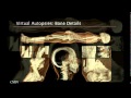
Now we have really made use of this, and we have tried to squeeze a lot of data into the system. And one of the applications that we've been working on -- and this has gotten a little bit of traction worldwide -- is the application of virtual autopsies. So again, looking at very, very large data sets, and you saw those full-body scans that we can do. We're just pushing the body through the whole CT scanner, and just in a few seconds we can get a full-body data set. So this is from a virtual autopsy. And you can see how I'm gradually peeling off. First you saw the body bag that the body came in, then I'm peeling off the skin -- you can see the muscles -- and eventually you can see the bone structure of this woman.
Now at this point, I would also like to emphasize that, with the greatest respect for the people that I'm now going to show -- I'm going to show you a few cases of virtual autopsies -- so it's with great respect for the people that have died under violent circumstances that I'm showing these pictures to you. In the forensic case -- and this is something that ... there's been approximately 400 cases so far just in the part of Sweden that I come from that has been undergoing virtual autopsies in the past four years. So this will be the typical workflow situation. The police will decide -- in the evening, when there's a case coming in -- they will decide, okay, is this a case where we need to do an autopsy? So in the morning, in between six and seven in the morning, the body is then transported inside of the body bag to our center and is being scanned through one of the CT scanners. And then the radiologist, together with the pathologist and sometimes the forensic scientist, looks at the data that's coming out, and they have a joint session. And then they decide what to do in the real physical autopsy after that.
Now looking at a few cases, here's one of the first cases that we had. You can really see the details of the data set. It's very high-resolution, and it's our algorithms that allow us to zoom in on all the details. And again, it's fully interactive, so you can rotate and you can look at things in real time on these systems here. Without saying too much about this case, this is a traffic accident, a drunk driver hit a woman. And it's very, very easy to see the damages on the bone structure. And the cause of death is the broken neck. And this women also ended up under the car, so she's quite badly beaten up by this injury.
Here's another case, a knifing. And this is also again showing us what we can do. It's very easy to look at metal artifacts that we can show inside of the body. You can also see some of the artifacts from the teeth -- that's actually the filling of the teeth -- but because I've set the functions to show me metal and make everything else transparent. Here's another violent case. This really didn't kill the person. The person was killed by stabs in the heart, but they just deposited the knife by putting it through one of the eyeballs. Here's another case. It's very interesting for us to be able to look at things like knife stabbings. Here you can see that knife went through the heart. It's very easy to see how air has been leaking from one part to another part, which is difficult to do in a normal, standard, physical autopsy. So it really, really helps the criminal investigation to establish the cause of death, and in some cases also directing the investigation in the right direction to find out who the killer really was.
Here's another case that I think is interesting. Here you can see a bullet that has lodged just next to the spine on this person. And what we've done is that we've turned the bullet into a light source, so that bullet is actually shining, and it makes it really easy to find these fragments. During a physical autopsy, if you actually have to dig through the body to find these fragments, that's actually quite hard to do.
One of the things that I'm really, really happy to be able to show you here today is our virtual autopsy table. It's a touch device that we have developed based on these algorithms, using standard graphics GPUs. It actually looks like this, just to give you a feeling for what it looks like. It really just works like a huge iPhone. So we've implemented all the gestures you can do on the table, and you can think of it as an enormous touch interface. So if you were thinking of buying an iPad, forget about it. This is what you want instead. Steve, I hope you're listening to this, all right. So it's a very nice little device. So if you have the opportunity, please try it out. It's really a hands-on experience. So it gained some traction, and we're trying to roll this out and trying to use it for educational purposes, but also, perhaps in the future, in a more clinical situation. There's a YouTube video that you can download and look at this, if you want to convey the information to other people about virtual autopsies.
Okay, now that we're talking about touch, let me move on to really "touching" data. And this is a bit of science fiction now, so we're moving into really the future. This is not really what the medical doctors are using right now, but I hope they will in the future. So what you're seeing on the left is a touch device. It's a little mechanical pen that has very, very fast step motors inside of the pen. And so I can generate a force feedback. So when I virtually touch data, it will generate forces in the pen, so I get a feedback. So in this particular situation, it's a scan of a living person. I have this pen, and I look at the data, and I move the pen towards the head, and all of a sudden I feel resistance. So I can feel the skin. If I push a little bit harder, I'll go through the skin, and I can feel the bone structure inside. If I push even harder, I'll go through the bone structure, especially close to the ear where the bone is very soft. And then I can feel the brain inside, and this will be the slushy like this.
So this is really nice. And to take that even further, this is a heart. And this is also due to these fantastic new scanners, that just in 0.3 seconds, I can scan the whole heart, and I can do that with time resolution. So just looking at this heart, I can play back a video here. And this is Karljohan,one of my graduate students who's been working on this project. And he's sitting there in front of the Haptic device, the force feedback system, and he's moving his pen towards the heart, and the heart is now beating in front of him, so he can see how the heart is beating. He's taken the pen, and he's moving it towards the heart, and he's putting it on the heart, and then he feels the heartbeats from the real living patient. Then he can examine how the heart is moving. He can go inside, push inside of the heart, and really feel how the valves are moving. And this, I think, is really the future for heart surgeons. I mean it's probably the wet dream for a heart surgeon to be able to go inside of the patient's heart before you actually do surgery, and do that with high-quality resolution data. So this is really neat.
Now we're going even further into science fiction. And we heard a little bit about functional MRI. Now this is really an interesting project. MRI is using magnetic fields and radio frequencies to scan the brain, or any part of the body. So what we're really getting out of this is information of the structure of the brain, but we can also measure the difference in magnetic properties of blood that's oxygenated and blood that's depleted of oxygen. That means that it's possible to map out the activity of the brain. So this is something that we've been working on. And you just saw Motts the research engineer, there, going into the MRI system, and he was wearing goggles. So he could actually see things in the goggles. So I could present things to him while he's in the scanner. And this is a little bit freaky, because what Motts is seeing is actually this. He's seeing his own brain. So Motts is doing something here, and probably he is going like this with his right hand, because the left side is activated on the motor cortex. And then he can see that at the same time. These visualizations are brand new. And this is something that we've been researching for a little while.
This is another sequence of Motts' brain. And here we asked Motts to calculate backwards from 100. So he's going "100,97,94." And then he's going backwards. And you can see how the little math processor is working up here in his brain and is lighting up the whole brain. Well this is fantastic. We can do this in real time. We can investigate things. We can tell him to do things. You can also see that his visual cortex is activated in the back of the head, because that's where he's seeing, he's seeing his own brain. And he's also hearing our instructions when we tell him to do things. The signal is really deep inside of the brain as well, and it's shining through, because all of the data is inside this volume. And in just a second here you will see -- okay, here. Motts, now move your left foot. So he's going like this. For 20 seconds he's going like that, and all of a sudden it lights up up here. So we've got motor cortex activation up there. So this is really, really nice, and I think this is a great tool. And connecting also with the previous talk here, this is something that we could use as a tool to really understand how the neurons are working, how the brain is working, and we can do this with very, very high visual quality and very fast resolution.
Now we're also having a bit of fun at the center. So this is a CAT scan -- Computer Aided Tomography. So this is a lion from the local zoo outside of Norrkoping in Kolmarden, Elsa. So she came to the center, and they sedated her and then put her straight into the scanner. And then, of course, I get the whole data set from the lion. And I can do very nice images like this. I can peel off the layer of the lion. I can look inside of it. And we've been experimenting with this. And I think this is a great application for the future of this technology, because there's very little known about the animal anatomy. What's known out there for veterinarians is kind of basic information. We can scan all sorts of things, all sorts of animals. The only problem is to fit it into the machine. So here's a bear. It was kind of hard to get it in. And the bear is a cuddly, friendly animal. And here it is. Here is the nose of the bear. And you might want to cuddle this one, until you change the functions and look at this. So be aware of the bear.
So with that, I'd like to thank all the people who have helped me to generate these images. It's a huge effort that goes into doing this, gathering the data and developing the algorithms, writing all the software. So, some very talented people. My motto is always, I only hire people that are smarter than I am and most of these are smarter than I am.
So thank you very much.
(Applause)
まず始めに医療に関係する データを取り扱う 課題について ご説明します これは私たちにとって大きな課題です そんな私たちを助けるのがこの機械です これはコンピュータ断層撮影装置 通称 CTです 素晴らしい装置です 人体の周りに対して 高速で回転するX線が使われます 機械を端から端まで通り抜けるのに およそ30秒かかり そこから膨大な 情報が出力されます 健康管理の向上のために 使える 大変素晴らしい機械です しかし 私たちにとっての課題でもあるのです その課題はこちらの画像で見て取れます これは現在私たちが直面している 医療データの爆発的な増加問題です この問題に現在取り組んでいます 過去に遡りましょう
数年前に何が起こったかご説明します これらの機械は 1970年頃に導入され始め 人体をスキャンし 100枚程度の人体画像を 生成します 大変勝手ながら 明確化するために それらをデータをデジタル化すると 大体50MBの大きさになります 情報量自体は私たちが 現在扱っているデータに比べると小さいでしょう 通常のモバイル機器で扱えます 電話帳に例えると 1メートル分積み上げた電話帳の数に相当します 現在私たちが扱っている これらの機械は 数秒以内に 2万4千枚もの人体の画像を生成します これは20GBものデータ もしくは 電話帳800冊分の情報量です 重ねれば200メートルは行くでしょう 何が起きようとしているか 今まさに起き始めている 技術トレンドとは 測定時間中の人体の状態を見れるようになったことです つまり人体の挙動を解剖なしで確認できるのです それでは 5秒もの間 データを取得したとしましょう それは1テラバイトものデータになります これは 80万冊の本 あるいは 16km分重ねた電話帳に相当します これは患者一人分のデータです 私たちが取り組む対象です
実に途方もない仕事です これで 2万5千枚もあります これらを放射線専門医が 対応する姿を想像してみてください 2万5千枚もの画像を前に こう言うのです 「えっと 2万5千枚か ああ ここが問題の箇所だ」 もうそのようなことはできません 不可能です そうするよりももっと合理的な方法を行う必要があります まずこれらの画像を一つにまとめます 自分自身の体をあらゆる角度から薄切りにし それらを再度元のデータの塊に戻すことを 想像してみてください そういうことを私たちは行っているのです このギガ テラバイト級のデータを元の塊に戻します もちろん データの塊は 単に 人体のそれぞれの箇所において 吸収されたX線の総量のみを表します そこで まずは私たちが 注目していないものは透過し 確認したいもののみを 見れるようにする必要があります つまり このような形に データを変換したいのです この課題は 私たちにとって とてつもない難題でした
絶えず処理速度や性能が良くなるコンピュータでも ギガバイトや テラバイト級のデータを対象に 関連情報を抽出するのは困難な作業です 心臓であったり血管や肝臓を 見たい時があるかもしれません もしかしたら腫瘍を発見する こともあるかもしれません そこでこの可愛らしい子の出番です これは私の娘です 今朝の9時頃の彼女の様子です 彼女はゲームで遊んでいます まだ2歳児ですが とても楽しんでいます 彼女はGPU開発の 推進役といっていいでしょう 子供達がゲームを遊ぶ限り グラフィックは進化し続けるのです 帰ったら 是非ゲームを勧めてください それが私たちには必要だからです
この機械の中には 私が医療データに対して行っていることを 可能にしてくれるものが存在します 実はこのような小さな装置を使っています ご存知のように おそらく10年程前 私が 1台目の画像処理用のコンピュータを 買う予算を得た時代の頃は それはとても巨大なマシンでした プロセッサやらストレージなどあらゆるものが詰め込まれていました 私はそのマシンに100万ドル支払いました そんな機械も 今や私のiPhoneと同じくらいの性能です 毎月 新型のグラフィックカードが販売されますが これは NVDIA ATI Intelといったベンダーから頂いた 数少ない最新モデルです ご存知のように 数百ドルも払えば このようなグラフィックカードを購入し コンピュータに追加して様々なことが可能になります このように これらと共に 人々に研究されている アルゴリズム開発や データの圧縮方法 関連情報の抽出方法などによって 膨大な医療データを取り扱うことを可能にしています
それでは 私たちが出来るいくつかの例をご紹介します これはCTスキャナーによって取得されたデータです ご覧のようにこれは完全なデータです これは女性で 髪の毛が見えます 女性の身体の個別構成を確認することができます このように歯の金属部分に対して X線が拡散していることが見て取れます ノイズが発生しているのはそういう部分です でも 通常のコンピュータ内の 標準のグラフィックカードによって インタラクティブに 断面を作ることができます 全てのデータは格納されているので 回転させたり 別の異なるアングルから確認ができます この女性は問題を抱えていたようですね 脳内出血を起こしていたようですが 小さなステントと呼ばれる血管を狭める 金属製の留め具で治療されています そして機能を変えることで 何を透明にし何を見えるようにするか 決めることができます 骨格を見ることができます これが 女性の頭蓋骨を開けた部分で ここから中に入ったようです これらは素晴らしい画像です 非常に解像度が高く 現在の標準グラフィックカードを使って 何が出来るかをとても良く示しています
効果的な利用方法を編み出した私たちは 膨大なデータをシステム上に 圧縮することを試みました 私たちの取り組んでいるアプリのうちの 一つは 世界中で少しずつ話題になりつつある バーチャル解剖アプリです 同じように 全身スキャンの画像といった 非常に巨大なデータセットを使います CTスキャナーに全身を通し 数秒後に全身の画像データを得ることができます これがバーチャル解剖です こうやって徐々に剥ぎ取っていきます 始めに遺体袋が確認できます 次に皮を剥ぎ取り 筋肉が見えますね 最後にこの女性の骨格がご覧になれます
ここからは これからお見せする人々に 対し敬意を持って いくつかのバーチャル解剖をお見せします 凶悪な事件によって お亡くなりになられた方々に敬意を払いつつ これらの画像を 法医学的な事例としてお見せします これらは私の出身である スウェーデンだけで 過去4年間において およそ400件バーチャル解剖が 執り行われています これが通常のワークフローです 警察は 例えば夕方に 事件が起きたとして 解剖の必要があるかどうか判断します そして朝の6時から7時にかけて 袋に包まれた遺体が 私たちのセンターに移送され CTスキャナーの一つでスキャンされます 病理学者やたまに法医学の研究者を 伴った放射線科医が 出力されるデータを確認し 合同で会議を開きます そして本当の解剖をどのように行うか決めるのです
いくつかの事例のうち 私たちが担当した初期の事例を紹介します このようにデータを本当に詳細に確認できます とても高画質です さらに私たちが考案したアルゴリズムによって 詳細な部分を拡大をすることが可能です 繰り返しますが 完全にインタラクティブで このシステムによってリアルタイムで 画像を回転させて見ることが可能です こちらの事例の説明は不要かもしれませんが これは交通事故です 酔っぱらった運転手が女性をひきました 骨格の損傷を確認することがとても容易です 死因は首の損傷です さらに女性は車に下敷きになってしまったため その怪我によって体に 重度の損傷を負っています
別の事例を紹介します 刺殺事件です 私たちにできることを この例でも説明します 体内に存在する金属製品を 簡単に確認することができます 歯の中の加工物も確認できます これは歯の詰め物です 金属のみを表示し その他は透明にする機能を 設定しています 別の凶悪な事例を紹介します これは実際の致命傷ではありません この方は心臓に複数回刺され 死亡しました しかし犯人はさらにナイフを 片方の眼球に刺したままにしたのです 別の事例を紹介します ナイフによる刺殺の事例などを検証できることは 私たちにとって大変興味深いものです ここではナイフが心臓に突き刺さっているのが確認できます 空気が一方からもう一方へと 漏れだしている様子が簡単に確認できます 通常の解剖ではこのようなことを確認することは困難です このように 死因を判断する上で 犯罪捜査に大変有効です また 捜査を正しい方向へ導き 真の殺人犯を 突き止める場合もあります
これは 私が興味深いと感じた事例です 弾丸がこの方の脊椎の横に 留まっているのが見えます この弾丸を光源に変換することで 弾丸がこれらの破片を光らせて 見つけやすくしています 通常の解剖を行う際にこれらの破片を 体内から見つけだそうとする場合は 実際には大変困難です
今日 皆さんにお見せ出来ることを 嬉しく思っているものの一つが このバーチャル解剖テーブルです これは標準のGPUと今までのアルゴリズムを 元に私たちが開発したタッチデバイスです 実際には このような形となっています 巨大なiPhoneのように動作します テーブル上で 行える全てのジェスチャーを実装しています 巨大なタッチインターフェースと考えて頂ければと思います iPadを買おうと考えている方は 忘れてください これこそ皆さんが欲していたものです スティーブ あなたがこれを聞いてくれているといいんですが という訳で これはとても良いデバイスです もし機会があれば 是非使ってみてください 実体験して頂くことをお勧めします 注目も集めたことなので 私たちはこれの教育目的の 利用を想定して製品化を目指しており 将来的には 医療現場での利用も考えています バーチャル解剖について紹介したい場合は YouTubeに閲覧できる動画があるので 是非ご利用ください
「触れる」ことについてご紹介したので 次は本当にデータに触れることについてお話します 若干SFが入ってきますが 未来のことについてお話します 今は 医師がこれを利用している訳ではありませんが 将来は 使っていることに期待しています 左側に見えるのはタッチデバイスです 小さな機械式のペンで 高速のステッピング・モーターが内蔵されており フィードバックを生むことができます よって データに仮想的に触れると ペンに接触力が生まれ 感覚を得ることができるのです このように生きている方の スキャン画像に対して ペンを持って データを確認しながら 頭部に向けてペンを動かすと 突如 抵抗力を感じることができます このように皮膚を感じることができます もう少し強く押すと 皮膚を通り抜け 中の骨格を感じることができます さらに強く押せば 骨格を通り抜け 耳のすぐ近くの柔らかい骨を通り ぬるぬるしたような感じで 脳の部分を感じることができます
非常に有効な機能です さらに例を紹介します これが心臓です 新型のスキャナーのおかげで たった0.3秒で 心臓全体をスキャンすることができます さらに時間分解が行えるため 心臓を見ながら 動画を再生することが可能です 彼はこのプロジェクトに取り組んでいる 大学院生の一人でカーデュアンと言います フィードバックシステムである触覚装置の前に座りながら 心臓に向けてペンを動かすと 心臓が目の前で拍動しはじめます どのように心臓が拍動するのか確認できるのです ペンをとって 心臓に向けて動かし 心臓の上にペンを置くと 生きた患者の心臓の鼓動を感じることができるため 心臓の動作を確認することができます 心臓の中に移動し 内部を押して 心臓弁の動作を感じることができます これこそが 心臓外科医の将来の姿であると考えます 心臓外科医にとって 患者の心臓の中を 高解像度のデータを元に手術前に 確認するなんて夢のような話でしょう 非常に素晴らしい構想です
さらにSFに近いものをご紹介します 機能MRIについてご存知でしょうか これはとても興味深いプロジェクトです MRIは磁場と周波数を利用し 脳や体の あらゆる部分をスキャンすることができます これによって 脳の構造についての情報が得られます しかし 更にこれを使って 酸素を含む血液と そうでない血液の磁性の差を測定することが可能です これはつまり 脳の活動を映し出すことが可能なのです 私たちはこれにも取り組んでいます ちょうど研究技術者であるモッツが MRIにゴーグルを着用して 中に入る所をご覧頂いています これはゴーグルを通して 彼にスキャナーにいながら映像を見せることができるためです これは中々ビックリするかもしれません モッツが実際に見ている映像はこれです 彼は自分の脳を見ているのです モッツはここで何かしていますね 恐らく右手でこういう風にしています なぜなら左側は運動皮質によって 活性化されるからです 彼も同じくその様子を確認できます このような可視化は新しい取り組みであり 私たちが少し前から研究している分野です
これはモッツの脳の別の部分です 彼には 100から逆に計算するように頼みました 「100 97 94...」とった具合に 計算しています 彼の脳の小さな計算に関わる領域が活性化し 脳の全体を光らせているのがわかります 素晴らしい結果です リアルタイムで計測できます 彼に依頼して調査を行うことが可能です 更に彼の視覚野が 頭頂部の後ろ側で活性化しています なぜなら自分自身の脳を見ているからです また彼は 私たちが彼に何かをさせるための 命令を聞いています この信号は脳の奥深くで発せられていますが 中で光っているのが確認できます 全てのデータがここに含まれているからです ここでは以下のような光景をご覧頂けます モッツ 左足を動かしてください 彼はその通りにします 20秒間そのままの状態でいます するとここが急に光ります 運動皮質が活性化されたことが確認できます 非常に面白い結果です これはとても素晴らしいツールだと思います そして今までお話しした内容をまとめると ニューロンや脳がどのように 機能しているのかを理解する上で とても使えるツールであると考えます 何より非常に高画質かつ高分解能な上 高速に処理できます
さらにセンター内で少々面白いことも行っています これはCAT(コンピューター断層撮影)スキャンです これはノーショーピングのはずれにある コルマルデン動物園からのエルサというライオンです 彼女はセンターに来て 鎮静状態にされ そのままスキャナー内に運び込みました その後 ライオンの全データを取得しました このようなライオンの画像に対して レイヤーを剥ぎ取っていき 内部を確認していきます このようにして検証を行ってきました これは未来のテクノロジーにおける 大変優れたアプリだと思います なぜなら 動物解剖学については未知の部分も多く 獣医側で知られているのは基本的な知識に限られています あらゆる動物をはじめ あらゆるものをスキャンできます 唯一の問題は機械の中に入れることくらいです これは熊です 機械に入れるのに苦労しました 熊は非常にかわいらしい 友好的な動物です これは 熊の鼻の部分です 抱きしめたくなるでしょう 機能を変更してこれを見るまでは 熊には注意しましょう
以上をもって これらの画像の生成を手伝って頂いた 全ての方々に感謝したいと思います データの収集やアルゴリズムの開発 全てのソフトウェアを作り上げるまでに 非常に多くの労力がかかっています 非常に能力のある方々のおかげです 私のモットーは 私よりも頭の良い人達を雇うことです 多くは私より頭が良い方々ばかりです
ありがとうございました
(拍手)
品詞分類
- 主語
- 動詞
- 助動詞
- 準動詞
- 関係詞等
TED 日本語
TED Talks
関連動画

うつやPTSDを予防できる新種の薬 | TED Talkレベッカ・ブラックマン
2019.04.17騒音が健康に有害な理由 ― そして私たちにできることマティアス・バスナー
2019.02.26さまよう心を鎮めるには | TED Talkアミシ・ジャー
2018.04.18鬱の友達と心を通わせるにはビル・バーナット
2018.03.23脳に良い変化をもたらす運動の効果ウェンディー・スズキ
おすすめ 12018.03.21物事の「良し悪し」は思い込みに過ぎないヘザー・ラニエ
おすすめ 12018.01.19脳が深い睡眠から更なる恩恵を得る方法ダン・ガーテンバーグ
2018.01.04うつを一人で抱え込まないでニッキー・ウェバー・アレン
2017.10.26長寿の秘訣は周囲の人との交流かもスーザン・ピンカー
2017.09.04脳はどのように美しさを判定するか?アンジャン・チャタジー
2017.08.22脳が「意識された現実」という幻覚を作り出す仕組みアニル・セス
2017.07.18注意を向けた時、脳では何が起きているのかメディ・オディカニ=セイドラー
2017.07.12ティーンエイジャーの登校時間を遅らせるべき理由とはウェンディ・トロクセル
2017.06.09死に直面したとき、人生に生きる価値を与えてくれるのはルーシー・カラニシ
おすすめ 12017.06.07最善の自己と最悪の自己の生物学ロバート・サポルスキー
2017.05.31メンタルヘルスを気遣うのは恥ずかしくなんかないサング・デリ
2017.05.26


洋楽 おすすめ
RECOMMENDS
洋楽歌詞
ダイナマイトビーティーエス
洋楽最新ヒット2020.08.20ディス・イズ・ミーグレイテスト・ショーマン・キャスト
洋楽人気動画2018.01.11グッド・ライフGイージー、ケラーニ
洋楽人気動画2017.01.27ホワット・ドゥ・ユー・ミーン?ジャスティン・ビーバー
洋楽人気動画2015.08.28ファイト・ソングレイチェル・プラッテン
洋楽人気動画2015.05.19ラヴ・ミー・ライク・ユー・ドゥエリー・ゴールディング
洋楽人気動画2015.01.22アップタウン・ファンクブルーノ・マーズ、マーク・ロンソン
洋楽人気動画2014.11.20ブレイク・フリーアリアナ・グランデ
洋楽人気動画2014.08.12ハッピーファレル・ウィリアムス
ポップス2014.01.08カウンティング・スターズワンリパブリック
ロック2013.05.31ア・サウザンド・イヤーズクリスティーナ・ペリー
洋楽人気動画2011.10.26ユー・レイズ・ミー・アップケルティック・ウーマン
洋楽人気動画2008.05.30ルーズ・ユアセルフエミネム
洋楽人気動画2008.02.21ドント・ノー・ホワイノラ・ジョーンズ
洋楽人気動画2008.02.15オンリー・タイムエンヤ
洋楽人気動画2007.10.03ミス・ア・シングエアロスミス
ロック2007.08.18タイム・トゥ・セイ・グッバイサラ・ブライトマン
洋楽人気動画2007.06.08シェイプ・オブ・マイ・ハートスティング
洋楽人気動画2007.03.18ウィ・アー・ザ・ワールド(U.S.A. フォー・アフリカ)マイケル・ジャクソン
洋楽人気動画2006.05.14ホテル・カリフォルニアイーグルス
ロック2005.07.06