TED日本語
TED Talks(英語 日本語字幕付き動画)
TED日本語 - ナンシー・カンウィッシャー: 脳神経が描く私達の思考
TED Talks
脳神経が描く私達の思考
A neural portrait of the human mind
ナンシー・カンウィッシャー
Nancy Kanwisher
内容
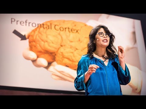
脳画像のパイオニア、ナンシー・カンウィッシャーは、fMRIで主に自分の脳の各領域で起こる活動の画像を撮り、彼女の同僚と共に研究した結果 ― 私たちの脳は、非常に特殊に分化した領域と、汎用の機能を持つ組織とで成っている ― という発見を、このトークで私達と共有します。驚く事に、それでも脳には未知の課題が多く残されているのです。


字幕
SCRIPT
Script

Today I want to tell you about a project being carried out by scientists all over the world to paint a neural portrait of the human mind. And the central idea of this work is that the human mind and brain is not a single, general-purpose processor, but a collection of highly specialized components, each solving a different specific problem, and yet collectively making up who we are as human beings and thinkers. To give you a feel for this idea,
imagine the following scenario: You walk into your child's day care center. As usual, there's a dozen kids there waiting to get picked up, but this time, the children's faces look weirdly similar, and you can't figure out which child is yours. Do you need new glasses? Are you losing your mind? You run through a quick mental checklist. No, you seem to be thinking clearly, and your vision is perfectly sharp. And everything looks normal except the children's faces. You can see the faces, but they don't look distinctive, and none of them looks familiar, and it's only by spotting an orange hair ribbon that you find your daughter.
This sudden loss of the ability to recognize faces actually happens to people. It's called prosopagnosia, and it results from damage to a particular part of the brain. The striking thing about it is that only face recognition is impaired; everything else is just fine.
Prosopagnosia is one of many surprisingly specific mental deficits that can happen after brain damage. These syndromes collectively have suggested for a long time that the mind is divvied up into distinct components, but the effort to discover those components has jumped to warp speed with the invention of brain imaging technology, especially MRI. So MRI enables you to see internal anatomy at high resolution, so I'm going to show you in a second a set of MRI cross-sectional images through a familiar object, and we're going to fly through them and you're going to try to figure out what the object is. Here we go.
It's not that easy. It's an artichoke.
Okay, let's try another one, starting from the bottom and going through the top. Broccoli! It's a head of broccoli. Isn't it beautiful? I love that.
Okay, here's another one. It's a brain, of course. In fact, it's my brain. We're going through slices through my head like that. That's my nose over on the right, and now we're going over here, right there.
So this picture's nice, if I do say so myself, but it shows only anatomy. The really cool advance with functional imaging happened when scientists figured out how to make pictures that show not just anatomy but activity, that is, where neurons are firing. So here's how this works. Brains are like muscles. When they get active, they need increased blood flow to supply that activity, and lucky for us, blood flow control to the brain is local, so if a bunch of neurons, say, right there get active and start firing, then blood flow increases just right there. So functional MRI picks up on that blood flow increase, producing a higher MRI response where neural activity goes up.
So to give you a concrete feel for how a functional MRI experiment goes and what you can learn from it and what you can't, let me describe one of the first studies I ever did. We wanted to know if there was a special part of the brain for recognizing faces, and there was already reason to think there might be such a thing based on this phenomenon of prosopagnosia that I described a moment ago, but nobody had ever seen that part of the brain in a normal person, so we set out to look for it. So I was the first subject. I went into the scanner, I lay on my back, I held my head as still as I could while staring at pictures of faces like these and objects like these and faces and objects for hours. So as somebody who has pretty close to the world record of total number of hours spent inside an MRI scanner, I can tell you that one of the skills that's really important for MRI research is bladder control. (Laughter)
When I got out of the scanner, I did a quick analysis of the data, looking for any parts of my brain that produced a higher response when I was looking at faces than when I was looking at objects, and here's what I saw. Now this image looks just awful by today's standards, but at the time I thought it was beautiful. What it shows is that region right there, that little blob, it's about the size of an olive and it's on the bottom surface of my brain about an inch straight in from right there. And what that part of my brain is doing is producing a higher MRI response, that is, higher neural activity, when I was looking at faces than when I was looking at objects. So that's pretty cool, but how do we know this isn't a fluke? Well, the easiest way is to just do the experiment again. So I got back in the scanner, I looked at more faces and I looked at more objects and I got a similar blob, and then I did it again and I did it again and again and again, and around about then I decided to believe it was for real. But still, maybe this is something weird about my brain and no one else has one of these things in there, so to find out, we scanned a bunch of other people and found that pretty much everyone has that little face-processing region in a similar neighborhood of the brain.
So the next question was, what does this thing really do? Is it really specialized just for face recognition? Well, maybe not, right? Maybe it responds not only to faces but to any body part. Maybe it responds to anything human or anything alive or anything round. The only way to be really sure that that region is specialized for face recognition is to rule out all of those hypotheses. So we spent much of the next couple of years scanning subjects while they looked at lots of different kinds of images, and we showed that that part of the brain responds strongly when you look at any images that are faces of any kind, and it responds much less strongly to any image you show that isn't a face, like some of these.
So have we finally nailed the case that this region is necessary for face recognition? No, we haven't. Brain imaging can never tell you if a region is necessary for anything. All you can do with brain imaging is watch regions turn on and off as people think different thoughts. To tell if a part of the brain is necessary for a mental function, you need to mess with it and see what happens, and normally we don't get to do that. But an amazing opportunity came about very recently when a couple of colleagues of mine tested this man who has epilepsy and who is shown here in his hospital bed where he's just had electrodes placed on the surface of his brain to identify the source of his seizures. So it turned out by total chance that two of the electrodes happened to be right on top of his face area. So with the patient's consent, the doctors asked him what happened when they electrically stimulated that part of his brain. Now, the patient doesn't know where those electrodes are, and he's never heard of the face area. So let's watch what happens. It's going to start with a control condition that will say "Sham" nearly invisibly in red in the lower left, when no current is delivered, and you'll hear the neurologist speaking to the patient first. So let's watch.
(Video) Neurologist: Okay, just look at my face and tell me what happens when I do this. All right?
Neurologist: One,two,three.
Patient: Nothing. Neurologist: Nothing? Okay. I'm going to do it one more time. Look at my face. One,two,three.
Patient: You just turned into somebody else. Your face metamorphosed. Your nose got saggy, it went to the left. You almost looked like somebody I'd seen before, but somebody different. That was a trip. (Laughter)
Nancy Kanwisher: So this experiment -- (Applause) -- this experiment finally nails the case that this region of the brain is not only selectively responsive to faces but causally involved in face perception. So I went through all of these details about the face region to show you what it takes to really establish that a part of the brain is selectively involved in a specific mental process. Next, I'll go through much more quickly some of the other specialized regions of the brain that we and others have found. So to do this, I've spent a lot of time in the scanner over the last month so I can show you these things in my brain.
So let's get started. Here's my right hemisphere. So we're oriented like that. You're looking at my head this way. Imagine taking the skull off and looking at the surface of the brain like that. Okay, now as you can see, the surface of the brain is all folded up. So that's not good. Stuff could be hidden in there. We want to see the whole thing, so let's inflate it so we can see the whole thing. Next, let's find that face area I've been talking about that responds to images like these. To see that, let's turn the brain around and look on the inside surface on the bottom, and there it is, that's my face area. Just to the right of that is another region that is shown in purple that responds when you process color information, and near those regions are other regions that are involved in perceiving places, like right now, I'm seeing this layout of space around me and these regions in green right there are really active. There's another one out on the outside surface again where there's a couple more face regions as well. Also in this vicinity is a region that's selectively involved in processing visual motion, like these moving dots here, and that's in yellow at the bottom of the brain, and near that is a region that responds when you look at images of bodies and body parts like these, and that region is shown in lime green at the bottom of the brain.
Now all these regions I've shown you so far are involved in specific aspects of visual perception. Do we also have specialized brain regions for other senses, like hearing? Yes, we do. So if we turn the brain around a little bit, here's a region in dark blue that we reported just a couple of months ago, and this region responds strongly when you hear sounds with pitch, like these. (Sirens) (Cello music) (Doorbell) In contrast, that same region does not respond strongly when you hear perfectly familiar sounds that don't have a clear pitch, like these. (Chomping) (Drum roll) (Toilet flushing)
Okay. Next to the pitch region is another set of regions that are selectively responsive when you hear the sounds of speech.
Okay, now let's look at these same regions. In my left hemisphere, there's a similar arrangement -- not identical, but similar -- and most of the same regions are in here, albeit sometimes different in size.
Now, everything I've shown you so far are regions that are involved in different aspects of perception, vision and hearing. Do we also have specialized brain regions for really fancy, complicated mental processes? Yes, we do. So here in pink are my language regions. So it's been known for a very long time that that general vicinity of the brain is involved in processing language, but we showed very recently that these pink regions respond extremely selectively. They respond when you understand the meaning of a sentence, but not when you do other complex mental things, like mental arithmetic or holding information in memory or appreciating the complex structure in a piece of music.
The most amazing region that's been found yet is this one right here in turquoise. This region responds when you think about what another person is thinking. So that may seem crazy, but actually, we humans do this all the time. You're doing this when you realize that your partner is going to be worried if you don't call home to say you're running late. I'm doing this with that region of my brain right now when I realize that you guys are probably now wondering about all that gray, uncharted territory in the brain, and what's up with that?
Well, I'm wondering about that too, and we're running a bunch of experiments in my lab right now to try to find a number of other possible specializations in the brain for other very specific mental functions. But importantly, I don't think we have specializations in the brain for every important mental function, even mental functions that may be critical for survival. In fact, a few years ago, there was a scientist in my lab who became quite convinced that he'd found a brain region for detecting food, and it responded really strongly in the scanner when people looked at images like this. And further, he found a similar response in more or less the same location in 10 out of 12 subjects. So he was pretty stoked, and he was running around the lab telling everyone that he was going to go on "Oprah" with his big discovery. But then he devised the critical test: He showed subjects images of food like this and compared them to images with very similar color and shape, but that weren't food, like these. And his region responded the same to both sets of images. So it wasn't a food area, it was just a region that liked colors and shapes. So much for "Oprah."
But then the question, of course, is, how do we process all this other stuff that we don't have specialized brain regions for? Well, I think the answer is that in addition to these highly specialized components that I've been describing, we also have a lot of very general- purpose machinery in our heads that enables us to tackle whatever problem comes along. In fact, we've shown recently that these regions here in white respond whenever you do any difficult mental task at all -- well, of the seven that we've tested. So each of the brain regions that I've described to you today is present in approximately the same location in every normal subject. I could take any of you, pop you in the scanner, and find each of those regions in your brain, and it would look a lot like my brain, although the regions would be slightly different in their exact location and in their size.
What's important to me about this work is not the particular locations of these brain regions, but the simple fact that we have selective, specific components of mind and brain in the first place. I mean, it could have been otherwise. The brain could have been a single, general-purpose processor, more like a kitchen knife than a Swiss Army knife. Instead, what brain imaging has delivered is this rich and interesting picture of the human mind. So we have this picture of very general-purpose machinery in our heads in addition to this surprising array of very specialized components.
It's early days in this enterprise. We've painted only the first brushstrokes in our neural portrait of the human mind. The most fundamental questions remain unanswered. So for example, what does each of these regions do exactly? Why do we need three face areas and three place areas, and what's the division of labor between them? Second, how are all these things connected in the brain? With diffusion imaging, you can trace bundles of neurons that connect to different parts of the brain, and with this method shown here, you can trace the connections of individual neurons in the brain, potentially someday giving us a wiring diagram of the entire human brain. Third, how does all of this very systematic structure get built, both over development in childhood and over the evolution of our species? To address questions like that, scientists are now scanning other species of animals, and they're also scanning human infants.
Many people justify the high cost of neuroscience research by pointing out that it may help us someday to treat brain disorders like Alzheimer's and autism. That's a hugely important goal, and I'd be thrilled if any of my work contributed to it, but fixing things that are broken in the world is not the only thing that's worth doing. The effort to understand the human mind and brain is worthwhile even if it never led to the treatment of a single disease. What could be more thrilling than to understand the fundamental mechanisms that underlie human experience, to understand, in essence, who we are? This is, I think, the greatest scientific quest of all time.
(Applause)
今日お話ししたい事は 世界中の科学者が行っている あるプロジェクト ヒトの脳神経が描く像についてです この研究の中心となる考えは ヒトの思考と脳は 汎用な単一目的の プロセッサーなのではなく 高度に特殊化された 各部の複合体であり その1つ1つが各々 固有の問題を解決しながらも 総合的に 思考する我々を 作っているという事です これを理解する為
次のシナリオを想像してみて下さい あなたは自分の子供がいる 保育所に入って行きます いつものように何十人もの子どもがいて 親の迎えを待っていますが 今日は 子ども達の顔が奇妙に同じに見えます どの子が自分の子なのか 見分けがつきません 新しい眼鏡がいるのでしょうか? 気がおかしくなったのでしょうか? 素早く色々な事が頭をよぎります いいえ頭ははっきりしていて 目も問題なく良く見えています 全てがまともなのに 子供達の顔だけがそうでないのです 顔は見えるのですが 区別ができず どの子も覚えのない顔なのです オレンジ色のリボンを見つけて ようやくそれが自分の子供だと分かります
このように急に顔を 認識できなくなることが 実際起きるのです それは相貌失認と呼ばれ 脳のある特定の場所に 障害が起きた結果起きます これに驚かされる事は 顔認知だけに障害があり その他は問題がない事です
相貌失認は脳損傷後におきる 数々の 驚くほど特異な 脳機能障害の1つです このような症状などを総合して 脳は明確に役割が分担された領域別に 分かれている事が長い間 示唆されていたのですが それ等の領域を探求する研究に 一気に進歩をもたらしたのは 脳画像技術に於ける発明 ― 特にMRIです MRIを使い脳内部の組織構造を 高解度画像で見ることができます MRIの断面画像を よくご存知のものを通して お見せしましょう その物とは何か分かるでしょうか これです
分かり難いですが アーティチョークです
ではこれはどうでしょう 下から上まで行きますよ ブロッコリーの頭です! 綺麗でしょう?私は好きです
もう1つ 勿論これはヒトの脳です 実はこれは私の脳です 私の脳をスライスして行きます 右の方に鼻があり 今ここに来ました ちょうどここです
この画像はいいですね 自分でも本当にそう思いますが 組織を映し出してるだけに過ぎません 機能画像という本当にすごい進歩は 科学者が組織だけでなく 活動 すなわちニューロンの発火を 捉える技術を 編み出した時に起きました それは このような働きをします 脳は筋肉の様なもので 活発な時は その活動の為に 送り込む血量を増やします 幸いに脳の血流制御は 局部的なので 例えばここで 多くのニューロンが 活発に発火し始めたら そこの血流は増します fMRIはその血流の 増加をキャッチし ニューロンが活性化した箇所で 高反応を示します
具体的に fMRIの実験が如何に行われるか そこからどんな事が分かるか 又 分からないかを説明するため 最初の研究の1つを お話しします 我々が知りたかったのは 顔認知だけの領野があるかどうかです 既に その存在は お話しした相貌失認に基づき 推察されてはいましたが 健常者の脳にそれを確認した者は 誰もいませんでした それで我々はその探索に 乗り出しました 私が最初の実験台になり MRI 内に入り横たわりました 頭を出来るだけ動かさず このような顔や物を 何時間も見つめました 私は fMRIスキャナーの中で 費やした時間の合計が 世界記録に 近いと思いますが この研究で 本当に大切なスキルの一つは 膀胱制御でした (笑)
スキャナーから出た時 データをすぐに分析して 私の脳の中で 顔を見ている時に 物体を見たときよりも 強い反応を示す箇所を探しました これが私が見たものです この画像は今の基準からすると ひどいもののように見えますが 当時は美しいものだと思いました 小さな塊が見える 箇所がありますが オリーブサイズ位で 私の脳表面の下部 ― ここから約2.5cm真っすぐ中に 入った所です 脳のその箇所が示しているのは 強い fMRI反応 つまり 非常に活発な脳神経の活動です 私が 物体でなく 人の顔写真を見ている時のものです かなり面白いのですが 何故これが偶然でないと 分かるのでしょう? では最も簡単な方法は 実験をもう一度やってみる事です 私はMRIスキャナーに戻り もっと顔や物の写真を見ました そして同じ様な斑点が見られました それからもう一度やり 又同じ結果がでました もう一度 もう一度と もうその頃には これは本物だと 信じる事にしました しかし これは私の脳が 何かおかしいからであり 他の誰にも こういう事は 起きないのかもしれないと 他の多くの人達も スキャンして調べると 殆ど同じ結果で 小さな顔領域が 誰でも 同じ様な部位にありました
それで次にくる疑問は この箇所の機能です 顔認知だけでしょうか? そうでないかもしれませんね 人の顔だけに反応するのでなく 他の身体部分にも 反応するかもしれません 人の様な物や他の生物や 丸っこい物にも 反応するかもしれません その領域が顔の認知に 特化して機能する事を はっきり確かめる唯一の方法は 仮説を全て除外していく事です そこで我々はそれから数年 色々な写真を見る人々の脳を スキャンして 何の顔であろうと 顔の写真を見ている時は 顔以外の写真の時と比べて 脳のその領野が 強く反応しているのが 観察されました
これで我々は ここは顔認知に必要な領域だと 決定づけたのでしょうか いいえ そうではありません 脳画像だけでは 特定の領野が 特定の機能に必要かどうかは 分からないのです 脳画像で私たちが出来る事は 様々な思考で あらゆる領野のニューロンが 活性・不活性化されるのを 見るだけです 脳の一部がある心的機能に 必要かどうかを見るには そこを刺激して 反応を見る必要がありますが 普段そういう事はできません しかし最近驚くべき機会が巡ってきて 私の同僚数人が 癲癇持ちのある男性を テストしました これが病院のベットに 横たわるその男性です 脳の表面に 癲癇の源を特定するための 電極が 配置されたばかりです 全く偶然に 電極の二つが 顔認知領域の上部に 配置されていました それで患者の同意を得て 医師達は 脳のその部位を電気で刺激して どうなるか彼に尋ねました 患者はどこに電極が あるのか知りませんし 顔認知領域の事など 聞いた事もありません では どうなるか見てみましょう 比較用の条件から始めます 殆ど見えないくらい「Sham」と 左下に赤く 電気が流されてない時に 出てきます 神経学者が患者にまず 話しかけているが聞こえます
神経学者:「私の顔を見て下さい こうした時に 何が起きるか言って下さい いいですか?」
患者:「OK」
神経学者:「1、2、3」
患者:「何も起きません」 神経学者:「何も?Ok」 神経学者:「もう一度します 私の顔を見て 1、2、3」
患者:「他の誰かになっただけです 顔が変形して 鼻が下がり左に行き 見た事がある様な 他の人みたいになりました でも全く他の誰かです ドラッグでもやったようでした」 (笑)
この実験が・・・ (拍手) この実験で ついに確定したことは この脳の領域は 顔だけに反応するのでなく 顔を認識する為に無くてはならないものだという事です こうして顔認知領域に関しての 詳細を見てきたのは 脳には特定の思考過程に関与する 領域がある事を確定するには どのようにする必要があるのかを お伝えする為です 次にもっと端的に 今までに発見された 機能が分化した脳領域 に関してお話しします この為に私は先月 fMRIの中で 時間を随分費やしたので 私の脳のこんなところを お見せできるのです
まず私の脳の右半球から行きます この向きから 今 私の頭をこのように見ています 頭蓋骨を取り除き 脳の表面を見ていると 想像してください ご覧のように 脳の表面が折り畳まれていますが 何か隠れているかもしれません 全部見たいので 膨らまして全体像を見ましょう お話ししてきたこのような写真に 反応する顔領域を 探しましょう 脳を回転して 下部の表面下を見てみましょう ここに私の顔領域があります その右には他の領域が 紫色に示してあり ここは色の情報を 処理する時 反応します その近くにはまた他の領域 場所を認識することに関わる領域があります 今私もこのように会場の空間レイアウトを 見ていますが ここにある緑色の領域は 本当に活発です もう1つ外側に 数カ所 顔認識領域があり この周辺にも 選択的に視覚運動機能に 関わっている領域があります この動いている 下部の黄色い斑点がそうです その近くには体の各部を 見る時に反応する領域があり その領域はライムグリーン色で 脳の下部にあります
これまでお見せした領域は 特定の視覚に関係しています また特定の機能 聴覚とか他の感覚を専門とする 領域があるでしょうか? あります 脳を少し回してみると 濃い青の領域があり ほんの数ヶ月前 発表したばかりです この領域は このような 音程の鮮明な音を聞くと 大きく反応します (サイレン) (チェロ音楽) (玄関の呼び鈴) それに比べ 同じ領域は こんな時は大きく反応しません 普段 よく聞く音 ― 次のように 音程が不鮮明な音を聞いた時です (咀嚼音) (ドラムの音) (トイレの水を流す音)
音程の鮮明さに反応する領域の側には こういう時だけに反応する 1つの領域帯があります 話し声を聞くときです
ではこれらの領域を見てみましょう 私の左半球は 同じ様になっていて 全く同じではありませんが ― 時にはサイズの 違いはあれ 領域の殆どがここにあります
これまでお見せした全ては 聴覚 視覚などの様々な 認知機能を司る 領域でした では 素晴らしく精巧で複雑な 思考活動のための 特別な領域もあるのでしょうか はい あります このピンクの色が私の言語領域です 長い間 脳の全般に於いて 言語プロセスは起きていると 思われてきました しかし ごく最近 このピンクの領域が 選択的に著しく 反応している事が分かりました センテンスの意味が分かる時 ここが反応しますが 他の複雑な思考には 反応しません それは暗算をするとか 情報を記憶しようとか 音楽の複雑な構成を理解しようと する時などがそうです
未だに分かっていない事で 最も驚くべき領域は 青緑色のこれです この領域は 他の人が考えている事を 推測している時に反応します クレイジーに聞こえますが 実際 我々はこんな事を 常にしています 遅くなる事を電話して 伝えなければパートナーが 心配するだろうと気づくとき あなたはこれをしています 今 私の脳がそれをしています というのも皆さんが多分 この触れてない灰色の部分は 一体何をするのだろうと 考えていらっしゃる事に 私は気がついているからです
私もそれについて考えていて 他に特定の機能を持った 領域が脳に多くあるのではないかと あらゆる実験を研究室で 行っています 重要な事は脳には 全ての思考機能それぞれに 致命的なものも含めて 特殊に特化した 機能が備わっているというのでは無い と思っています 事実 数年前 私の実験室に 食物を見分ける 領域を発見したと主張する 科学者がいました 人々がこのような画像を見たとき その領域はfMRIで大きく反応しました 更に実験は進められ 同じ様な反応が 殆ど同じ領域に12件中 10件の対象について 見られました それで彼はかなり興奮して 実験室を走り回り 「この大発見で『Oprah』に出るぞ」と みんなに言っていましたが 臨界実験を考えつき この様な食べ物の画像を被験者に見せ 色も形もよく似ているけれど 食べ物ではない このような物の画像と 比べて見せたところ その領域はその両方ともに 同じように反応したのです 食物領域ではなかったのです 色と形に反応するだけの 領域だったのです 『Oprah』は諦めるしかないですね
それでも 勿論 疑問が残ります 特化した脳領域が無い 他の全ての事をどうやって 脳は処理するのでしょうか その答えは お話しして来た 高度に分化した構成要素に加えて あらゆる問題に対処する 汎用な目的を持った 領域もあります 事実 白い領域は どんな難しい思考活動にも ― 少なくとも私達の7回の実験に於いて ― 反応する事が最近示されました 今日お話しした脳の各領域は 健常な人の脳には ほぼ同じ箇所にあります あなた方をMRIの スキャナーに入れ 脳にこれらの領域 1つ1つを探してみると この私の脳とよく似ているでしょう ただサイズや正確な位置には わずかな違いはあるでしょうが
この研究で私にとって重要な事は これらの領野がどこにあるかでなく 我々の脳には特殊に分化した 思考機能の構成分野が 初めからあったという シンプルな事実です つまり さもなければ 脳は単一の汎用機能を 有していただけでしょう スイスナイフというより 料理用のナイフの様なものです そうではなく 脳画像が示すのは 興味深い豊かなヒトの 思考活動を表しています 我々の頭の中には 汎用な目的を持つ 脳機能と 驚くほど複雑に特化され 多岐に分化した領域とが 共に存在するのです
この研究は始まったばかりで 精神活動のニューロン像を 描き始めたばかりに過ぎず 最も根本的な疑問は そのまま残っています 例えばこれらの領域は それぞれ何をしているのでしょうか? どうして3カ所も顔認知や 位置認知に必要なのでしょうか? これらの働きの分担は どうなっているのでしょうか? 第二に脳内部の それぞれの領域は どう関連し合っているのでしょうか? 拡散画像で 脳のあらゆる部分を繋ぐ ニューロンの束を辿ったり ここで示した方法で 脳のニューロン個別の繋がりを 辿る事が出来 こうして いつか脳全体の配線図が 分かる事となるでしょう 第三にこの様な体系的な構造が どうやって ヒトの成長期や人類の進化上 出来上がったのかです この様な疑問に取り組むため 科学者達は他の動物達の脳を スキャンして また幼児の脳もスキャンして 調べています
神経科学研究にかかる高額な 費用の必要性については アルツハイマー病や自閉症の様な 脳疾患の治療の為に いつか役に立つのだ と多くの人が指摘します それはとても重要な目標です 私の仕事がそう貢献できるなら どんなに嬉しいでしょう しかし 壊れた物を治す事だけが 唯一 価値があるのではありません ヒトの思考と脳を理解する努力は 病気を1つも 治せないとしても 価値のある事です 我々が本質的に誰なのかを 理解する為に ヒトの経験の根底となる 根本的機能を理解する事程 興奮させられる事はあるでしょうか これは科学に於ける 時代を超えた 最大の探求なのです
(拍手)
品詞分類
- 主語
- 動詞
- 助動詞
- 準動詞
- 関係詞等
TED 日本語
TED Talks
関連動画

うつやPTSDを予防できる新種の薬 | TED Talkレベッカ・ブラックマン
2019.04.17騒音が健康に有害な理由 ― そして私たちにできることマティアス・バスナー
2019.02.26さまよう心を鎮めるには | TED Talkアミシ・ジャー
2018.04.18鬱の友達と心を通わせるにはビル・バーナット
2018.03.23脳に良い変化をもたらす運動の効果ウェンディー・スズキ
おすすめ 12018.03.21物事の「良し悪し」は思い込みに過ぎないヘザー・ラニエ
おすすめ 12018.01.19脳が深い睡眠から更なる恩恵を得る方法ダン・ガーテンバーグ
2018.01.04うつを一人で抱え込まないでニッキー・ウェバー・アレン
2017.10.26長寿の秘訣は周囲の人との交流かもスーザン・ピンカー
2017.09.04脳はどのように美しさを判定するか?アンジャン・チャタジー
2017.08.22脳が「意識された現実」という幻覚を作り出す仕組みアニル・セス
2017.07.18注意を向けた時、脳では何が起きているのかメディ・オディカニ=セイドラー
2017.07.12ティーンエイジャーの登校時間を遅らせるべき理由とはウェンディ・トロクセル
2017.06.09死に直面したとき、人生に生きる価値を与えてくれるのはルーシー・カラニシ
おすすめ 12017.06.07最善の自己と最悪の自己の生物学ロバート・サポルスキー
2017.05.31メンタルヘルスを気遣うのは恥ずかしくなんかないサング・デリ
2017.05.26


洋楽 おすすめ
RECOMMENDS
洋楽歌詞
ステイザ・キッド・ラロイ、ジャスティン・ビーバー
洋楽最新ヒット2021.08.20スピーチレス~心の声ナオミ・スコット
洋楽最新ヒット2019.05.23シェイプ・オブ・ユーエド・シーラン
洋楽人気動画2017.01.30フェイデッドアラン・ウォーカー
洋楽人気動画2015.12.03ウェイティング・フォー・ラヴアヴィーチー
洋楽人気動画2015.06.26シー・ユー・アゲインウィズ・カリファ
洋楽人気動画2015.04.06シュガーマルーン5
洋楽人気動画2015.01.14シェイク・イット・オフテイラー・スウィフト
ポップス2014.08.18オール・アバウト・ザット・ベースメーガン・トレイナー
ポップス2014.06.11ストーリー・オブ・マイ・ライフワン・ダイレクション
洋楽人気動画2013.11.03コール・ミー・メイビーカーリー・レイ・ジェプセン
洋楽人気動画2012.03.01美しき生命コールドプレイ
洋楽人気動画2008.08.04バッド・デイ~ついてない日の応援歌ダニエル・パウター
洋楽人気動画2008.05.14サウザンド・マイルズヴァネッサ・カールトン
洋楽人気動画2008.02.19イッツ・マイ・ライフボン・ジョヴィ
ロック2007.10.11アイ・ウォント・イット・ザット・ウェイバックストリート・ボーイズ
洋楽人気動画2007.09.14マイ・ハート・ウィル・ゴー・オンセリーヌ・ディオン
洋楽人気動画2007.07.12ヒーローマライア・キャリー
洋楽人気動画2007.03.21オールウェイズ・ラヴ・ユーホイットニー・ヒューストン
洋楽人気動画2007.02.19オネスティビリー・ジョエル
洋楽人気動画2005.09.16