TED日本語
TED Talks(英語 日本語字幕付き動画)
TED日本語 - キエン・グエン: 色分けされた手術
TED Talks
色分けされた手術
Color-coded surgery
キエン・グエン
Quyen Nguyen
内容
細胞の種類ごとに色分けされた教科書と、実際の手術は今まで全く異なるものでしたが、これからは違います。TEDMEDではキエン・グエンが分子マーカーを使って、どこを切ればいいのかが見えるように腫瘍を緑色に光らせます。


字幕
SCRIPT
Script
I want to talk to you about one of the biggest myths in medicine, and that is the idea that all we need are more medical breakthroughs and then all of our problems will be solved. Our society loves to romanticize the idea of the single, solo inventor who, working late in the lab one night, makes an earthshaking discovery, and voila, overnight everything's changed. That's a very appealing picture, however, it's just not true. In fact, medicine today is a team sport. And in many ways, it always has been. I'd like to share with you a story about how I've experienced this very dramatically in my own work.
I'm a surgeon, and we surgeons have always had this special relationship with light. When I make an incision inside a patient's body, it's dark. We need to shine light to see what we're doing. And this is why, traditionally, surgeries have always started so early in the morning -- to take advantage of daylight hours. And if you look at historical pictures of the early operating rooms, they have been on top of buildings. For example, this is the oldest operating room in the Western world, in London, where the operating room is actually on top of a church with a skylight coming in. And then this is a picture of one of the most famous hospitals in America. This is Mass General in Boston. And do you know where the operating room is? Here it is on the top of the building with plenty of windows to let light in.
So nowadays in the operating room, we no longer need to use sunlight. And because we no longer need to use sunlight, we have very specialized lights that are made for the operating room. We have an opportunity to bring in other kinds of lights -- lights that can allow us to see what we currently don't see. And this is what I think is the magic of fluorescence.
So let me back up a little bit. When we are in medical school, we learn our anatomy from illustrations such as this where everything's color-coded. Nerves are yellow, arteries are red, veins are blue. That's so easy anybody could become a surgeon, right? However, when we have a real patient on the table, this is the same neck dissection -- not so easy to tell the difference between different structures. We heard over the last couple days what an urgent problem cancer still is in our society, what a pressing need it is for us to not have one person die every minute. Well if cancer can be caught early, enough such that someone can have their cancer taken out, excised with surgery, I don't care if it has this gene or that gene, or if it has this protein or that protein, it's in the jar. It's done, it's out, you're cured of cancer.
This is how we excise cancers. We do our best, based upon our training and the way the cancer looks and the way it feels and its relationship to other structures and all of our experience, we say, you know what, the cancer's gone. We've made a good job. We've taken it out. That's what the surgeon is saying in the operating room when the patient's on the table. But then we actually don't know that it's all out. We actually have to take samples from the surgical bed, what's left behind in the patient, and then send those bits to the pathology lab. In the meanwhile, the patient's on the operating room table. The nurses, anesthesiologist, the surgeon, all the assistants are waiting around. And we wait. The pathologist takes that sample, freezes it, cuts it, looks in the microscope one by one and then calls back into the room. And that may be 20 minutes later per piece. So if you've sent three specimens, it's an hour later. And very often they say, "You know what, points A and B are okay, but point C, you still have some residual cancer there. Please go cut that piece out." So we go back and we do that again, and again.
And this whole process: "Okay you're done. We think the entire tumor is out." But very often several days later, the patient's gone home, we get a phone call: "I'm sorry, once we looked at the final pathology, once we looked at the final specimen, we actually found that there's a couple other spots where the margins are positive. There's still cancer in your patient." So now you're faced with telling your patient, first of all, that they may need another surgery, or that they need additional therapy such as radiation or chemotherapy. So wouldn't it be better if we could really tell, if the surgeon could really tell, whether or not there's still cancer on the surgical field? I mean, in many ways, the way that we're doing it, we're still operating in the dark.
So in 2004, during my surgical residency, I had the great fortune to meet Dr. Roger Chen, who went on to win the Nobel Prize for chemistry in 2008. Roger and his team were working on a way to detect cancer, and they had a very clever molecule that they had come up with. The molecule they had developed had three parts. The main part of it is the blue part, polycation, and it's basically very sticky to every tissue in your body.
So imagine that you make a solution full of this sticky material and inject it into the veins of someone who has cancer, everything's going to get lit up. Nothing will be specific. There's no specificity there. So they added two additional components. The first one is a polyanionic segment, which basically acts as a non-stick backing like the back of a sticker. So when those two are together, the molecule is neutral and nothing gets stuck down. And the two pieces are then linked by something that can only be cut if you have the right molecular scissors -- for example, the kind of protease enzymes that tumors make. So here in this situation, if you make a solution full of this three-part molecule along with the dye, which is shown in green, and you inject it into the vein of someone who has cancer, normal tissue can't cut it. The molecule passes through and gets excreted. However, in the presence of the tumor, now there are molecular scissors that can break this molecule apart right there at the cleavable site. And now, boom, the tumor labels itself and it gets fluorescent.
So here's an example of a nerve that has tumor surrounding it. Can you tell where the tumor is? I couldn't when I was working on this. But here it is. It's fluorescent. Now it's green. See, so every single one in the audience now can tell where the cancer is. We can tell in the operating room, in the field, at a molecular level, where is the cancer and what the surgeon needs to do and how much more work they need to do to cut that out. And the cool thing about fluorescence is that it's not only bright, it actually can shine through tissue. The light that the fluorescence emits can go through tissue. So even if the tumor is not right on the surface, you'll still be able to see it.
In this movie, you can see that the tumor is green. There's actually normal muscle on top of it. See that? And I'm peeling that muscle away. But even before I peel that muscle away, you saw that there was a tumor underneath. So that's the beauty of having a tumor that's labeled with fluorescent molecules. That you can, not only see the margins right there on a molecular level, but you can see it even if it's not right on the top -- even if it's beyond your field of view. And this works for metastatic lymph nodes also.
Sentinel lymph node dissection has really changed the way that we manage breast cancer, melanoma. Women used to get really debilitating surgeries to excise all of the axillary lymph nodes. But when sentinel lymph node came into our treatment protocol, the surgeon basically looks for the single node that is the first draining lymph node of the cancer. And then if that node has cancer, the woman would go on to get the axillary lymph node dissection. So what that means is if the lymph node did not have cancer, the woman would be saved from having unnecessary surgery.
But sentinel lymph node, the way that we do it today, is kind of like having a road map just to know where to go. So if you're driving on the freeway and you want to know where's the next gas station, you have a map to tell you that that gas station is down the road. It doesn't tell you whether or not the gas station has gas. You have to cut it out, bring it back home, cut it up, look inside and say, "Oh yes, it does have gas." So that takes more time. Patients are still on the operating room table. Anesthesiologists, surgeons are waiting around. That takes time.
So with our technology, we can tell right away. You see a lot of little, roundish bumps there. Some of these are swollen lymph nodes that look a little larger than others. Who amongst us hasn't had swollen lymph nodes with a cold? That doesn't mean that there's cancer inside. Well with our technology, the surgeon is able to tell immediately which nodes have cancer. I won't go into this very much, but our technology, besides being able to tag tumor and metastatic lymph nodes with fluorescence, we can also use the same smart three-part molecule to tag gadolinium onto the system so you can do this noninvasively. The patient has cancer, you want to know if the lymph nodes have cancer even before you go in. Well you can see this on an MRI.
So in surgery, it's important to know what to cut out. But equally important is to preserve things that are important for function. So it's very important to avoid inadvertent injury. And what I'm talking about are nerves. Nerves, if they are injured, can cause paralysis, can cause pain. In the setting of prostate cancer, up to 60 percent of men after prostate cancer surgery may have urinary incontinence and erectile disfunction. That's a lot of people to have a lot of problems -- and this is even in so-called nerve-sparing surgery, which means that the surgeon is aware of the problem, and they are trying to avoid the nerves.
But you know what, these little nerves are so small, in the context of prostate cancer, that they are actually never seen. They are traced just by their known anatomical path along vasculature. And they're known because somebody has decided to study them, which means that we're still learning about where they are. Crazy to think that we're having surgery, we're trying to excise cancer, we don't know where the cancer is. We're trying to preserve nerves; we can't see where they are.
So I said, wouldn't it be great if we could find a way to see nerves with fluorescence? And at first this didn't get a lot of support. People said, "We've been doing it this way for all these years. What's the problem? We haven't had that many complications." But I went ahead anyway. And Roger helped me. And he brought his whole team with him. So there's that teamwork thing again. And we eventually discovered molecules that were specifically labeling nerves. And when we made a solution of this, tagged with the fluorescence and injected in the body of a mouse, their nerves literally glowed. You can see where they are.
Here you're looking at a sciatic nerve of a mouse, and you can see that that big, fat portion you can see very easily. But in fact, at the tip of that where I'm dissecting now, there's actually very fine arborizations that can't really be seen. You see what looks like little Medusa heads coming out. We have been able to see nerves for facial expression, for facial movement, for breathing -- every single nerve -- nerves for urinary function around the prostate. We've been able to see every single nerve. When we put these two probes together ... So here's a tumor. Do you guys know where the margins of this tumor is? Now you do. What about the nerve that's going into this tumor? That white portion there is easy to see. But what about the part that goes into the tumor? Do you know where it's going? Now you do.
Basically, we've come up with a way to stain tissue and color-code the surgical field. This was a bit of a breakthrough. I think that it'll change the way that we do surgery. We published our results in the proceedings of the National Academy of Sciences and in Nature Biotechnology. We received commentary in Discover magazine, in The Economist. And we showed it to a lot of my surgical colleagues. They said, "Wow! I have patients who would benefit from this. I think that this will result in my surgeries with a better outcome and fewer complications."
What needs to happen now is further development of our technology along with development of the instrumentation that allows us to see this sort of fluorescence in the operating room. The eventual goal is that we'll get this into patients. However, we've discovered that there's actually no straightforward mechanism to develop a molecule for one-time use. Understandably, the majority of the medical industry is focused on multiple-use drugs, such as long-term daily medications. We are focused on making this technology better. We're focused on adding drugs, adding growth factors, killing nerves that are causing problems and not the surrounding tissue. We know that this can be done and we're committed to doing it.
I'd like to leave you with this final thought. Successful innovation is not a single breakthrough. It is not a sprint. It is not an event for the solo runner. Successful innovation is a team sport, it's a relay race. It requires one team for the breakthrough and another team to get the breakthrough accepted and adopted. And this takes the long-term steady courage of the day-in day-out struggle to educate, to persuade and to win acceptance. And that is the light that I want to shine on health and medicine today.
Thank you very much.
(Applause)
今日お話したいことは 医学の 大きな誤った妄想の一つについてです それは 医学的な 大発見を続けさえすれば 私たちの抱える 問題を全て解決してくれるという考えです 私達の社会は 夢みがちです 研究している科学者が 独りきりで ある時夜遅くに 世界を揺るがすような発見をする ジャーン! 一夜にして全てが変わってしまう とても魅力的な想像です しかし実際はそうではありません 現実では 医学は団体競技なのです 様々な意味で ずっとそうでした 私が実際にそれを まざまざと体験したときのことを お話ししましょう
私は外科医です そして私たち外科医は常に 光と密接な関係がありました (光あれ!)患者の身体の中を切開するときはとても暗いのです 何をしているかの確認に光が必要です そのため 手術は伝統的に 朝早くに開始して 日光を有効活用しているのです この昔の絵を見ると 初期の手術室が大体 建物の一番上に置かれていたのが分かります 西洋で一番古い手術室が ロンドンにあるこの手術室です 手術室は 教会の一番上にあり 日光を取り入れるようにしています そしてこれはアメリカでもっとも有名な 病院のひとつである ボストンのマサチューセッツ総合病院です 手術室はどこでしょう? まさにここ 建物の頂上にあり たくさんの窓から光が入るようになっています
今日の手術室では 日光は必要なくなりました 日光を使う替わりに 手術室用に作られた 照明器具を使うからです 肉眼で見える光ではなく 今までは見えなかったものが 見えるようになる 違う種類の光をもたらすことができます これが私の考えている光 蛍光の魔法です
少し補足させてください 医学部での授業では 画面のように図解の解剖図で学び 全てが色分けされていました 神経は黄色 動脈は赤色 そして静脈は青色です 誰でも外科医になれそうですよね しかし これは首の図と同じ部位ですが 実際の手術で目にするとき それぞれの組織を見分けるのは そう簡単ではありません ここ数日間で 私たちは ガンが社会的にまだまだ 緊急問題であることと 毎分1人が がんで 死なざるを得ない状況の解決策が 強く求められていることを聞いてきました ガンが早期に見つかり 手術によって摘出することが可能だったのであれば そのガン細胞に どんな 遺伝子やタンパク質が 含まれていても関係ありません もうビンの中にあるのですから 摘出は完了し ガンが治療されたのです
ガン切除法をお話ししします 医者は研修やガンの見え方 触診や他の組織との繋がり 経験の全てを以て 最善を尽くします 医者は言います よしガンはなくなった 成し遂げた 摘出した 患者を手術状態としたままで こんな風に外科医は話します 実際には全て摘出したかまだわからないからです 手術台の患者の切除後の 周辺数か所から検体をとり 病理検査室へ送る必要があります その検査の間 患者は手術台の上にいて 看護師 麻酔医 外科医 そして 助手は待っているのです 待つしかないのです 病理医はその検体を 凍らせ 切断し 一つずつ顕微鏡で確認し 手術室へ結果を伝えます 一つにつき20分かかります もし3つ検体があったら 1時間かかるのです そして病理医が言うのは '実は AとBは大丈夫ですが" 'Cにはまだガンが残っているので" 'その部分を切除してください" 手術ではこの検査と切除を何回も続けます
そして次のように言うのです "終わりました" '全ての腫瘍は摘出されたようです" 数日後に患者は帰宅しますが 私たち担当医に電話がかかり '残念ですが' '最終の病理検査を" '最後の検体に行っていたところ" 'ガンの陽性反応がでました" '患者にはまだガンが残っています" と告げられるのは とてもよくある話です こうなると医者は患者に伝える事態に直面します まずは 再手術もしくは 放射線療法や化学療法などの 追加の治療が必要なことを伝えるのです 外科医が手術中に 術野にガンが残っているかを 確実に分かる方が 良いに決まっていますよね 様々な意味で手術のやり方は まだ暗闇の中で手術するようなものなのです
2004年 外科医の研修中に ロジャー・チェン先生と 出会う幸運に恵まれました 2008年にノーベル化学賞を 受賞した方です ロジャーとそのチームは ガンの発見方法を研究しており とても賢い分子を 開発していました 彼らが開発した分子は 3つの部分からできています 主要な部分は青い部分 ポリカチオンで どの体細胞にも とてもくっつきやすいものです
この成分だけを 使ったものを ガン患者に投薬したとしても 全てが光ってしまいます これでは特定できません まったく特異性がないのです そこで彼らはもう2つの成分を加えました 1つ目は赤い部分 ポリアニオンで シールの裏側のように くっつかない役目を果たします この2つを一緒にすると 分子は中性になり 体細胞にくっつかなくなります 2つ目は黄色部分で2つを繋ぐ役目を果たします 黄色部分は決められたハサミでしか切れません 例えば腫瘍が作り出す プロテアーゼ酵素を ハサミに設定します 次のように使います この3部構成の分子で溶液を作って 緑色で表されている 染料と一緒にガン患者の静脈に 投薬します すると 通常の細胞はハサミとならず 分子は体内を通り排泄されます しかし腫瘍があった場合 腫瘍がハサミとなり この分子を腫瘍部で 切断します そして こうなります 腫瘍は標識化され 蛍光に光ります
これは腫瘍が神経の周りに できている例です 腫瘍がどこかわかりますか? 私のときはわかりませんでした しかし ほら 光るのです 緑色の部分が見えます そう 今なら皆さん全員が ガンの位置が分かりますね 手術室 現場でも分子レベルでガンの場所や 外科医が摘出のために どう切除すれば良いのか どの程度周りを切除するのか 分かるのです また 蛍光のすごいところは 明るいだけではなく 組織の裏からでも光るところです 蛍光が発する光は 組織を 通り抜けられるのです なので 腫瘍が表面になかったとしても 見ることができるのです
この動画では 腫瘍が 緑色になっているのが見えます 実は正常な筋肉が手前にあります 見えますか? その筋肉をどかしてみましょう しかし筋肉をめくる前でも 腫瘍が裏側にあるのが見えました これが腫瘍を蛍光で 標識化することの長所なのです 境目が分子レベルで 見えるだけではなく 表面ではない通常見えないところに あったとしても見えるのです またリンパ節転移発見にも活用できます
センチネルリンパ節切除は 乳がんやメラノーマの治療を大きく変えました 以前までは腋下のリンパ節を 全て切除するという 患者を弱らせてしまう手術が必要でした センチネルリンパ節切除が標準的な 手術方法となった今 外科医は リンパ節でもガンに浸潤された 筋だけを探せばよくなりました もしその節にガンがあった場合 その女性は腋下リンパ節の 切除手術を受けるのです つまり リンパ節にガンが見つからなかった場合 その女性は不必要な 手術を受ける必要がないのです
しかし現在の方法ではセンチネルリンパ節は どこへ行くかを示す ただの地図に過ぎません 例えば高速道路を運転していて 次のガソリンスタンドを知りたければ 道の先にガソリンスタンドがあることがわかります しかしそこにガソリンが あるかどうかまではわかりません 切り出して 家にもって帰り スライスして 中を見て やっと ガソリンがあることがわかるのです このやり方だと多くの時間が必要です 検査の間も患者はまだ手術台にいます 麻酔医や外科医は待っています 時間がかかるのです
私たちの技術を使えばその場でわかります 小さな丸いコブが見えますね この中に肥大したリンパ節があります 他のものと比べて少しだけ大きいものです 風邪でも肥大したリンパ節は現れます 肥大してもガンがあるとは限らないのです 私たちの技術があれば 外科医はすぐさま どこにガンがあるか見分けられます 深くはお話しませんが 私たちの技術は 腫瘍や転移性リンパ節に蛍光で標識化するだけではなく 同じ3部構成の分子を使って 切ったりすることなく ガドリニウムの標識化もできます ガン患者に対して リンパ節がガンに侵されているかを 手術する前に確認できます MRIで見ることができます
手術では何を切除するのかを 知るのが重要です しかし同様に 体機能に大切なものを 温存することも重要です 意図しない損傷を避けることがとても重要です ここで私がお話しているのは 神経のことです 神経は傷つけられると 麻痺や 痛みを引き起こします 前立腺がんを例に取ると 60%の男性が 前立腺がんの手術のあと 尿漏れと勃起不全を 起こしています 多くの人に多くの問題が起こってしまうのです そしてこれは 外科医が十分に注意して 神経を避けるような手術 神経温存手術でさえ 起こっているのです
前立腺がんの場合には これらの神経はとても小さく 決して見ないのです 血管構造に沿っていると知られている 解剖学的な神経経路を たどっているに過ぎません これは誰かが研究しているからこそわかることですが それは実際の場所は まだ研究中ということです こんな手術で大丈夫でしょうか ガンの場所がわからないのに切除しようとしているのです 神経の場所がわからないのに温存しようとしているのです
お伝えしたように 蛍光によって 神経を標識化できれば すばらしくないですか? 最初はあまり支援されませんでした 人には"ずっとこのやり方をしてきましたよ 問題ないですよ 合併症もそれほどないよ" と言われました しかし私は突き進みました ロジャーが助けてくれたのです チーム全てを引き連れてきてくれました ここでも団体競技の必要性がわかります そしてついに神経を標識化する 分子を見つけたのです これで溶液を作り 蛍光で標識を付け マウスに投与したら 神経が文字通り輝いていたのです どこが神経かわかります
これはマウスの坐骨神経です 大きく太い部分がよく分かるでしょう しかし 私が今指し示しているところには とても微細な分枝がありますが 見えません 小さいメデューサの頭のようなものが見えますね 顔の表情や動き 呼吸 それらの全ての神経 前立腺のあたりの排尿の神経も 見ることができます 神経一つずつを見ることができるのです この二つが指し示していることをお教えしましょう ここに腫瘍があります この腫瘍の縁がわかりますか? これでわかりますね この腫瘍につながっている神経はどうでしょう? 白い部分は見やすいですが 腫瘍の中に入っている部分はどうでしょう? どこに入っているかわかりますか? これでわかりますね
基本的には 私達は医療現場で 細胞を染色し 色分けをする方法を開発したのです これはちょっとした革命です 手術のあり方を変えると思っています 私達は研究結果を 米国科学アカデミー紀要と ネイチャー バイオテクノロジーで 発表しました ディスカバリーとエコノミストでは 解説を頂いています 多くの同僚の外科医に見せたところ "すごい 私の患者も 恩恵を受けられるだろう この方法なら 私の手術は より良い結果が出せて 合併症も少なくなる" と言っていました
今 待ち望まれているのは この技術の更なる発達と 通常の手術室で この蛍光標識を 可能にする仕組みの 開発です 最終的なゴールは これらを患者に適用することです しかし 患者各々に特有の 蛍光分子を開発するのは 単純にできるものではないと わかりました 当然ながら医薬業界では 皆が使える長期にわたる日用服用薬などに 焦点が当てられています 私達はこの技術を発展させることに専念しています また新薬や成長因子を発展させたり 周囲の細胞を傷つけずに 原因の神経を殺傷したり することに注力しています 私達はこれが可能であること やるべきことであると信じています
最後にこの考えを皆さんに伝えます 本当の革新というのは 一つのひらめきだけでは成し得ません 短距離走ではありません 個人競技ではないのです 本当の革新というのは 団体競技のリレーなのです 一つのチームがひらめきを生み もうひとつのチームが それを適応させ浸透させるのです そのためには長期にわたって昼夜を問わずに 知らしめ説得し 認知を得る たゆまぬ勇気が 必要なのです 今日の健康と医療に投じたいのは この光なのです
どうもありがとう
(拍手)
品詞分類
- 主語
- 動詞
- 助動詞
- 準動詞
- 関係詞等
TED 日本語
TED Talks
関連動画

うつやPTSDを予防できる新種の薬 | TED Talkレベッカ・ブラックマン
2019.04.17騒音が健康に有害な理由 ― そして私たちにできることマティアス・バスナー
2019.02.26さまよう心を鎮めるには | TED Talkアミシ・ジャー
2018.04.18鬱の友達と心を通わせるにはビル・バーナット
2018.03.23脳に良い変化をもたらす運動の効果ウェンディー・スズキ
おすすめ 12018.03.21物事の「良し悪し」は思い込みに過ぎないヘザー・ラニエ
おすすめ 12018.01.19脳が深い睡眠から更なる恩恵を得る方法ダン・ガーテンバーグ
2018.01.04うつを一人で抱え込まないでニッキー・ウェバー・アレン
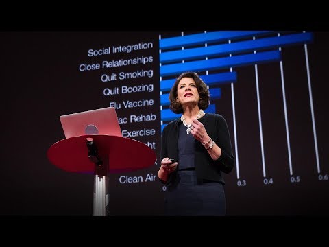
2017.10.26長寿の秘訣は周囲の人との交流かもスーザン・ピンカー
2017.09.04脳はどのように美しさを判定するか?アンジャン・チャタジー
2017.08.22脳が「意識された現実」という幻覚を作り出す仕組みアニル・セス
2017.07.18注意を向けた時、脳では何が起きているのかメディ・オディカニ=セイドラー
2017.07.12ティーンエイジャーの登校時間を遅らせるべき理由とはウェンディ・トロクセル
2017.06.09死に直面したとき、人生に生きる価値を与えてくれるのはルーシー・カラニシ
おすすめ 12017.06.07最善の自己と最悪の自己の生物学ロバート・サポルスキー
2017.05.31メンタルヘルスを気遣うのは恥ずかしくなんかないサング・デリ
2017.05.26

洋楽 おすすめ
RECOMMENDS
洋楽歌詞
ステイザ・キッド・ラロイ、ジャスティン・ビーバー
洋楽最新ヒット2021.08.20スピーチレス~心の声ナオミ・スコット
洋楽最新ヒット2019.05.23シェイプ・オブ・ユーエド・シーラン
洋楽人気動画2017.01.30フェイデッドアラン・ウォーカー
洋楽人気動画2015.12.03ウェイティング・フォー・ラヴアヴィーチー
洋楽人気動画2015.06.26シー・ユー・アゲインウィズ・カリファ
洋楽人気動画2015.04.06シュガーマルーン5
洋楽人気動画2015.01.14シェイク・イット・オフテイラー・スウィフト
ポップス2014.08.18オール・アバウト・ザット・ベースメーガン・トレイナー
ポップス2014.06.11ストーリー・オブ・マイ・ライフワン・ダイレクション
洋楽人気動画2013.11.03コール・ミー・メイビーカーリー・レイ・ジェプセン
洋楽人気動画2012.03.01美しき生命コールドプレイ
洋楽人気動画2008.08.04バッド・デイ~ついてない日の応援歌ダニエル・パウター
洋楽人気動画2008.05.14サウザンド・マイルズヴァネッサ・カールトン
洋楽人気動画2008.02.19イッツ・マイ・ライフボン・ジョヴィ
ロック2007.10.11アイ・ウォント・イット・ザット・ウェイバックストリート・ボーイズ
洋楽人気動画2007.09.14マイ・ハート・ウィル・ゴー・オンセリーヌ・ディオン
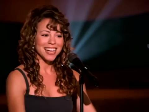
洋楽人気動画2007.07.12ヒーローマライア・キャリー
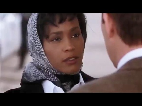
洋楽人気動画2007.03.21オールウェイズ・ラヴ・ユーホイットニー・ヒューストン
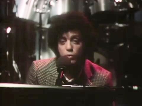
洋楽人気動画2007.02.19オネスティビリー・ジョエル
洋楽人気動画2005.09.16