TED日本語
TED Talks(英語 日本語字幕付き動画)
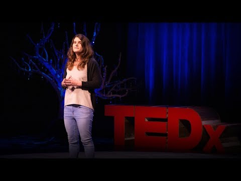
TED日本語 - ドリュー・ベリー: 不可視な超微小生物世界のCG
TED Talks
不可視な超微小生物世界のCG
Animations of unseeable biology
ドリュー・ベリー
Drew Berry
内容
分子や分子の動作を直接観察する方法はありません。ドリュー・ベリーはこれを変えるため、研究者がこれまで観察できなかった細胞内で起こる変化の過程をCGにしました。TEDxSydney で紹介する アニメーションは科学的に正確なだけでなく見る人を楽しませてくれます。
字幕
SCRIPT
Script

What I'm going to show you are the astonishing molecular machines that create the living fabric of your body. Now molecules are really, really tiny. And by tiny, I mean really. They're smaller than a wavelength of light, so we have no way to directly observe them. But through science, we do have a fairly good idea of what's going on down at the molecular scale. So what we can do is actually tell you about the molecules, but we don't really have a direct way of showing you the molecules.
One way around this is to draw pictures. And this idea is actually nothing new. Scientists have always created pictures as part of their thinking and discovery process. They draw pictures of what they're observing with their eyes, through technology like telescopes and microscopes, and also what they're thinking about in their minds. I picked two well-known examples, because they're very well-known for expressing science through art.
And I start with Galileo who used the world's first telescope to look at the Moon. And he transformed our understanding of the Moon. The perception in the 17th century was the Moon was a perfect heavenly sphere. But what Galileo saw was a rocky, barren world, which he expressed through his watercolor painting.
Another scientist with very big ideas, the superstar of biology, is Charles Darwin. And with this famous entry in his notebook, he begins in the top left-hand corner with, "I think," and then sketches out the first tree of life, which is his perception of how all the species, all living things on Earth, are connected through evolutionary history -- the origin of species through natural selection and divergence from an ancestral population.
Even as a scientist, I used to go to lectures by molecular biologists and find them completely incomprehensible, with all the fancy technical language and jargon that they would use in describing their work, until I encountered the artworks of David Goodsell, who is a molecular biologist at the Scripps Institute. And his pictures, everything's accurate and it's all to scale. And his work illuminated for me what the molecular world inside us is like.
So this is a transection through blood. In the top left-hand corner, you've got this yellow-green area. The yellow-green area is the fluids of blood, which is mostly water, but it's also antibodies, sugars, hormones, that kind of thing. And the red region is a slice into a red blood cell. And those red molecules are hemoglobin. They are actually red; that's what gives blood its color. And hemoglobin acts as a molecular sponge to soak up the oxygen in your lungs and then carry it to other parts of the body.
I was very much inspired by this image many years ago, and I wondered whether we could use computer graphics to represent the molecular world. What would it look like? And that's how I really began. So let's begin.
This is DNA in its classic double helix form. And it's from X-ray crystallography, so it's an accurate model of DNA. If we unwind the double helix and unzip the two strands, you see these things that look like teeth. Those are the letters of genetic code, the 25,000 genes you've got written in your DNA. This is what they typically talk about -- the genetic code -- this is what they're talking about. But I want to talk about a different aspect of DNA science, and that is the physical nature of DNA. It's these two strands that run in opposite directions for reasons I can't go into right now. But they physically run in opposite directions, which creates a number of complications for your living cells, as you're about to see, most particularly when DNA is being copied.
And so what I'm about to show you is an accurate representation of the actual DNA replication machine that's occurring right now inside your body, at least 2002 biology. So DNA's entering the production line from the left-hand side, and it hits this collection, these miniature biochemical machines, that are pulling apart the DNA strand and making an exact copy. So DNA comes in and hits this blue, doughnut-shaped structure and it's ripped apart into its two strands. One strand can be copied directly, and you can see these things spooling off to the bottom there. But things aren't so simple for the other strand because it must be copied backwards. So it's thrown out repeatedly in these loops and copied one section at a time, creating two new DNA molecules.
Now you have billions of this machine right now working away inside you, copying your DNA with exquisite fidelity. It's an accurate representation, and it's pretty much at the correct speed for what is occurring inside you. I've left out error correction and a bunch of other things. This was work from a number of years ago. Thank you.
This is work from a number of years ago, but what I'll show you next is updated science, it's updated technology. So again, we begin with DNA. And it's jiggling and wiggling there because of the surrounding soup of molecules, which I've stripped away so you can see something. DNA is about two nanometers across, which is really quite tiny. But in each one of your cells, each strand of DNA is about 30 to 40 million nanometers long. So to keep the DNA organized and regulate access to the genetic code, it's wrapped around these purple proteins -- or I've labeled them purple here. It's packaged up and bundled up. All this field of view is a single strand of DNA. This huge package of DNA is called a chromosome. And we'll come back to chromosomes in a minute.
We're pulling out, we're zooming out, out through a nuclear pore, which is the gateway to this compartment that holds all the DNA called the nucleus. All of this field of view is about a semester's worth of biology, and I've got seven minutes. So we're not going to be able to do that today? No, I'm being told, "No."
This is the way a living cell looks down a light microscope. And it's been filmed under time-lapse, which is why you can see it moving. The nuclear envelope breaks down. These sausage-shaped things are the chromosomes, and we'll focus on them. They go through this very striking motion that is focused on these little red spots. When the cell feels it's ready to go, it rips apart the chromosome. One set of DNA goes to one side, the other side gets the other set of DNA -- identical copies of DNA. And then the cell splits down the middle. And again, you have billions of cells undergoing this process right now inside of you.
Now we're going to rewind and just focus on the chromosomes and look at its structure and describe it. So again, here we are at that equator moment. The chromosomes line up. And if we isolate just one chromosome, we're going to pull it out and have a look at its structure. So this is one of the biggest molecular structures that you have, at least as far as we've discovered so far inside of us. So this is a single chromosome. And you have two strands of DNA in each chromosome. One is bundled up into one sausage. The other strand is bundled up into the other sausage.
These things that look like whiskers that are sticking out from either side are the dynamic scaffolding of the cell. They're called mircrotubules. That name's not important. But what we're going to focus on is this red region -- I've labeled it red here -- and it's the interface between the dynamic scaffolding and the chromosomes. It is obviously central to the movement of the chromosomes. We have no idea really as to how it's achieving that movement.
We've been studying this thing they call the kinetochore for over a hundred years with intense study, and we're still just beginning to discover what it's all about. It is made up of about 200 different types of proteins, thousands of proteins in total. It is a signal broadcasting system. It broadcasts through chemical signals telling the rest of the cell when it's ready, when it feels that everything is aligned and ready to go for the separation of the chromosomes. It is able to couple onto the growing and shrinking microtubules.
It's involved with the growing of the microtubules, and it's able to transiently couple onto them. It's also an attention sensing system. It's able to feel when the cell is ready, when the chromosome is correctly positioned. It's turning green here because it feels that everything is just right. And you'll see, there's this one little last bit that's still remaining red. And it's walked away down the microtubules. That is the signal broadcasting system sending out the stop signal. And it's walked away. I mean, it's that mechanical. It's molecular clockwork.
This is how you work at the molecular scale. So with a little bit of molecular eye candy, we've got kinesins, which are the orange ones. They're little molecular courier molecules walking one way. And here are the dynein. They're carrying that broadcasting system. And they've got their long legs so they can step around obstacles and so on. So again, this is all derived accurately from the science. The problem is we can't show it to you any other way.
Exploring at the frontier of science, at the frontier of human understanding, is mind-blowing. Discovering this stuff is certainly a pleasurable incentive to work in science. But most medical researchers -- discovering the stuff is simply steps along the path to the big goals, which are to eradicate disease, to eliminate the suffering and the misery that disease causes and to lift people out of poverty.
Thank you.
(Applause)

皆さんにお見せするのは 人体の生体組織を造っている 驚くべき分子マシンです 分子は非常に非常に小さいのです 本当に かなり小さいのです 光の波長より小さいので 直接見ることはできません でも科学のおかげで小さな分子の世界で 何が起きているのか かなり分かっています しかし分子についてお話する事はできても お見せする直接の方法はありません
見えないものを 絵で表現するという方法は 決して目新しいものではありません 科学者達はこれまでも 考えや発見の段階で絵を使ってきました 望遠鏡や顕微鏡を覗いて見た事や 頭の中で考えている事を 絵に描きました アートで科学を表現する という点で有名な2つの例をご紹介します
まずはガリレオ 世界初の望遠鏡で 月をみた人物ですよね 月の知識を一変させました 17世紀当時 月は完璧な 美しい球体だとされていましたが ガリレオが見たのはゴツゴツした不毛なもので 彼はそれを水彩画で表現しました
もう一人は チャールズ・ダーウィンです 壮大な考えを持っていた生物学界のスターです この有名なスケッチの左上には 「私の考えでは」とありそれから 最初の生命の樹が描かれています 地球上の全生物が 進化過程でどう繋がっているか という彼の説を表しています 祖先からの多様化と 自然淘汰による生物種の起源が表現されています
ところで 科学者の私でさえ 分子生物学の講義を受けては 研究の説明に専門用語や特殊用語が頻出し 内容が全く理解できない と感じることが よくありました そんな時分子生物学者デイヴィッド・グッドセルの 美術作品に出会いました 彼の絵は 構造も縮尺も 正確に表現されています 体内の分子世界がどうなっているのかが 彼の作品では理解できました
例えば 血液の断面図です 左上の端の黄緑色のエリアがありますね これは血液の流体でほとんどが水ですが 免疫体 糖 ホルモン等を 含んでいます 赤色の所は赤血球の断面で 赤い分子はヘモグロビンです 血液が赤いのはこのためです ヘモグロビンは分子のスポンジの役割をし 酸素を肺で吸収し 体全体に運びます
私は何年も前に この絵に刺激され コンピューターグラフィックを用いて 分子の世界を表現できないか考えました どう見えるだろうなぁって そこから始めたんです ではいきますよ
ご存知の2重螺旋のDNA こちらX線解析によるもので 正確なDNAのモデルです 螺旋をばらして2つの鎖を解くと 歯のようなものが現れます これは遺伝子コードの文字列で 25,000のヒトの遺伝子をDNA上に書いています 遺伝子コードってよく耳にしますね ご覧のこれがまさにそれなんです ここではDNA科学の違った側面_ DNAの物理的な性質をお話します これは逆向きに並んでいる2本の鎖です 細かい理由は省きますが 鎖の方向性が逆になっているため 私たちの細胞にとって不便なことが起こります ご覧になればわかりますが 特にDNAの複写時に面倒なことが起こります
次の画像は 今まさに 皆さんの体内でも活動している DNA複製機の正確なモデルです 2002年時点の生物学ですが DNAが左側から生産ラインに入って行き、 二本のDNAをバラバラにし 全く同じコピーを作る 超小型 生物化学装置に達します DNAが入ってきて ドーナツ型の青い部分にあたると 鎖は2本に引き裂かれます 片方の鎖は直接複写され 下の方へ巻き落ちて行きますが もう片方の鎖では そう単純にはいきません 前後逆に複製する必要があるからです 繰り返し この様なループにされ 一部ごと複写されて 2セットの二本鎖DNA分子が造られます
今こうしている間もあなたの体内で 何十億個ものこの機械が活動し 精巧かつ完全な複製を作っています 正確に表現できています 複写速度も ほぼこの速さです ここでは エラー修正や他の様々なことは省略しています ここまでは数年前の作品です ありがとうございます
これは随分前のものでしたが 今からお見せするのは 新しい科学知識を さらに進んだ技術で表現したものです 今回も DNAから始めましょう 通常は分子を含んだ液体の中で振動していますが 見やすいように液体を取り除きました DNAの幅は約2ナノメートルで とても小さいのですが 私たちの細胞内のDNAは 3千から4千ナノメートルの長さがあります DNAをまとめ 遺伝子コードへのアクセスを制限するために タンパク質が周りを包んでいます ここでは紫色で現されています 包まれて束ねられています 画面全体に広がるのはたった1本のDNAなんですよ この巨大なDNAの包みが染色体です 染色体については後でお話しするとして
ズームアウトして 全DNAを含む 細胞核という所から 核膜孔を抜けて 出て見ましょう ちなみに映っているものは 生物のクラス 一学期分に値しますが 7分しかないので 今日は全部お話できませんね? 「駄目」だそうです
光学顕微鏡で覗くと細胞はこう見えます 低速度撮影のため動くのが見えています 核膜が破れました ソーセージのような形のが染色体で ここを中心に見ていきます 染色体が著しい動きをしている 箇所が赤い部分に集中しています 細胞分裂の準備が整うと 染色体は2つに分かれ 一組のDNAセットは一方へ もう一組は他方へ行きます 複製した全く同じDNAです そして細胞が真ん中で分離します 繰り返しますが今も体内では 何十億という細胞がこうして分裂しています
では少し巻き戻して 染色体だけに着目して 構造を見て 解説しましょう 分裂の瞬間に戻ってきました 染色体が並んでいます 1つの染色体を取り出して 構造を見てみましょう これは現在の生物学上 体内で 最も大きい分子構造の1つです これが1つの染色体で 分裂期の染色体には2つのDNAの鎖が入っています 一方は1つのソーセージに 他方は別のに束ねられています
ひげのような物が両側に突き出しているのが見えますね ここは微小管といいます 細胞分裂の足場になります 名前は重要ではありません 赤く色付けられた所に注目しましょう ここは 伸び縮みする足場と 染色体の結合部です 明らかに 染色体の動きの中枢です この動きの仕組みは はっきり解っていません
これは動原体と呼ばれて かなり綿密に 100年以上研究されてきましたが その働きが やっと少しずつ解ってきたところです 合計数千個にも及ぶ約200種類もの タンパク質から出来ています 動原体は 信号伝達のシステムです 全てが揃って準備ができると 細胞の他の部分に 染色体が切り離せる状態であることを 化学的な信号で知らせます 動原体は伸縮する微小管に結合する働きもします
それは微小管を伸ばすと同時に 一時的に結合することも出来ます これは検知システムでもあり 細胞が準備できた時や染色体が 正しく並べられた時が分かります 全てが準備できると ここが緑色に変わります ここに 小さく1つだけ 赤色のままのものが あります 赤色部分は微小管を歩いて離れていきます これは伝達システムが「停止」の信号を送っているのです 歩いて離れる まさに機械的な動作です 細かく正確な動きです
このように分子の世界は動いています ちょっと見た目が面白い分子に オレンジ色のキネシンがあります 小さな分子の運び屋で左に進んでいます これはダイニンで 伝達システムを担っています ダイニンは長い脚で障害物をかわしたりします これは科学から得られた情報を 正確に画像としたもので 視覚的に説明する唯一の方法です他の方法では見ることができません
最先端の科学や 最先端の 人類の知識を探求することは 強烈で刺激的なものです このような発見が 科学者の原動力になっていることは確かです しかし 殆どの医療研究者にとって このような発見をすることは 大きな目標への通過点でしかありません 大きな目標は病気を撲滅し 病気からの苦しみや悲しみをなくし 貧困をなくす事です
ありがとうございました
(拍手)
品詞分類
- 主語
- 動詞
- 助動詞
- 準動詞
- 関係詞等
TED 日本語
TED Talks
関連動画

プラスチックを食べる細菌モーガン・ヴェイグ
2019.06.24
微生物の世界へようこそ ― 自宅にも、そしてあなたの顔にもアン・マデン
2017.08.24
CRISPRについて、みんなが知るべきことエレン・ヨルゲンセン
2016.10.24
自然に潜む驚異的な力を生かす方法オーデッド・ショゼヨフ
2016.10.18
自分でDNA検査ができる時代の到来セバスチャン・クレイベス
2016.10.13
リンゴから耳を作るマッドサイエンティストアンドリュー・ペリング
2016.07.08
1つの生物種全体を永久に変えてしまう遺伝子編集技術ジェニファー・カーン
2016.06.02
ゲノムを読んで人間を作る方法リッカルド・サバティーニ
2016.05.24
宇宙での生存に備えて人類が進化する方法リサ・ニップ
2016.04.21
干ばつに耐えられる農作物の作り方ジル・ファラント
2016.02.09
DNA編集が可能な時代、使い方は慎重にジェニファー・ダウドナ
2015.11.12
テクノロジーとバイオロジーを融合したデザインネリ・オックスマン
2015.10.29
アニメーションで科学者の仮説を試す方法ジャネット・イワサ
2014.08.07
二人の若き科学者、バクテリアでプラスチックを分解
2013.07.18
毛長マンモスを復活させよう!ヘンドリック・ポイナー
2013.05.30
私たちを取り巻く細菌と住環境のデザインジェシカ・グリーン
2013.03.25
洋楽 おすすめ
RECOMMENDS
洋楽歌詞

ダイナマイトビーティーエス
洋楽最新ヒット2020.08.20
ディス・イズ・ミーグレイテスト・ショーマン・キャスト
洋楽人気動画2018.01.11
グッド・ライフGイージー、ケラーニ
洋楽人気動画2017.01.27
ホワット・ドゥ・ユー・ミーン?ジャスティン・ビーバー
洋楽人気動画2015.08.28
ファイト・ソングレイチェル・プラッテン
洋楽人気動画2015.05.19
ラヴ・ミー・ライク・ユー・ドゥエリー・ゴールディング
洋楽人気動画2015.01.22
アップタウン・ファンクブルーノ・マーズ、マーク・ロンソン
洋楽人気動画2014.11.20
ブレイク・フリーアリアナ・グランデ
洋楽人気動画2014.08.12
ハッピーファレル・ウィリアムス
ポップス2014.01.08
カウンティング・スターズワンリパブリック
ロック2013.05.31
ア・サウザンド・イヤーズクリスティーナ・ペリー
洋楽人気動画2011.10.26
ユー・レイズ・ミー・アップケルティック・ウーマン
洋楽人気動画2008.05.30
ルーズ・ユアセルフエミネム
洋楽人気動画2008.02.21
ドント・ノー・ホワイノラ・ジョーンズ
洋楽人気動画2008.02.15
オンリー・タイムエンヤ
洋楽人気動画2007.10.03
ミス・ア・シングエアロスミス
ロック2007.08.18
タイム・トゥ・セイ・グッバイサラ・ブライトマン
洋楽人気動画2007.06.08
シェイプ・オブ・マイ・ハートスティング
洋楽人気動画2007.03.18
ウィ・アー・ザ・ワールド(U.S.A. フォー・アフリカ)マイケル・ジャクソン
洋楽人気動画2006.05.14
ホテル・カリフォルニアイーグルス
ロック2005.07.06


