TED日本語
TED Talks(英語 日本語字幕付き動画)
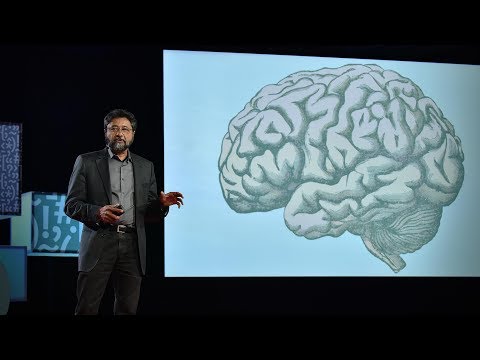
TED日本語 - カール・シューノーヴァー: 脳の中身を見る方法
TED Talks
脳の中身を見る方法
How to look inside the brain
カール・シューノーヴァー
Carl Schoonover
内容
脳の研究における進歩には 目を見張るものがあるが、実際どうやって脳内のニューロンを調べるのか?神経科学者でTEDフェローのカール・シューノーヴァーが、魅力的な写真の数々を使いながら、脳の中身を見せてくれるツールを紹介します。


字幕
SCRIPT
Script
This is a thousand-year-old drawing of the brain. It's a diagram of the visual system. And some things look very familiar today. Two eyes at the bottom, optic nerve flowing out from the back. There's a very large nose that doesn't seem to be connected to anything in particular.
And if we compare this to more recent representations of the visual system, you'll see that things have gotten substantially more complicated over the intervening thousand years. And that's because today we can see what's inside of the brain, rather than just looking at its overall shape.
Imagine you wanted to understand how a computer works and all you could see was a keyboard, a mouse, a screen. You really would be kind of out of luck. You want to be able to open it up, crack it open, look at the wiring inside. And up until a little more than a century ago, nobody was able to do that with the brain. Nobody had had a glimpse of the brain's wiring.
And that's because if you take a brain out of the skull and you cut a thin slice of it, put it under even a very powerful microscope, there's nothing there. It's gray, formless. There's no structure. It won't tell you anything.
And this all changed in the late 19th century. Suddenly, new chemical stains for brain tissue were developed and they gave us our first glimpses at brain wiring. The computer was cracked open.
So what really launched modern neuroscience was a stain called the Golgi stain. And it works in a very particular way. Instead of staining all of the cells inside of a tissue, it somehow only stains about one percent of them. It clears the forest, reveals the trees inside. If everything had been labeled, nothing would have been visible. So somehow it shows what's there.
Spanish neuroanatomist Santiago Ramon y Cajal, who's widely considered the father of modern neuroscience, applied this Golgi stain, which yields data which looks like this, and really gave us the modern notion of the nerve cell, the neuron. And if you're thinking of the brain as a computer, this is the transistor. And very quickly Cajal realized that neurons don't operate alone, but rather make connections with others that form circuits just like in a computer. Today, a century later, when researchers want to visualize neurons, they light them up from the inside rather than darkening them. And there's several ways of doing this. But one of the most popular ones involves green fluorescent protein. Now green fluorescent protein, which oddly enough comes from a bioluminescent jellyfish, is very useful. Because if you can get the gene for green fluorescent protein and deliver it to a cell, that cell will glow green -- or any of the many variants now of green fluorescent protein, you get a cell to glow many different colors.
And so coming back to the brain, this is from a genetically engineered mouse called "Brainbow." And it's so called, of course, because all of these neurons are glowing different colors.
Now sometimes neuroscientists need to identify individual molecular components of neurons, molecules, rather than the entire cell. And there's several ways of doing this, but one of the most popular ones involves using antibodies. And you're familiar, of course, with antibodies as the henchmen of the immune system. But it turns out that they're so useful to the immune system because they can recognize specific molecules, like, for example, the code protein of a virus that's invading the body. And researchers have used this fact in order to recognize specific molecules inside of the brain, recognize specific substructures of the cell and identify them individually.
And a lot of the images I've been showing you here are very beautiful, but they're also very powerful. They have great explanatory power. This, for example, is an antibody staining against serotonin transporters in a slice of mouse brain.
And you've heard of serotonin, of course, in the context of diseases like depression and anxiety. You've heard of SSRIs, which are drugs that are used to treat these diseases. And in order to understand how serotonin works, it's critical to understand where the serontonin machinery is. And antibody stainings like this one can be used to understand that sort of question.
I'd like to leave you with the following thought: Green fluorescent protein and antibodies are both totally natural products at the get-go. They were evolved by nature in order to get a jellyfish to glow green for whatever reason, or in order to detect the code protein of an invading virus, for example. And only much later did scientists come onto the scene and say, "Hey, these are tools, these are functions that we could use in our own research tool palette." And instead of applying feeble human minds to designing these tools from scratch, there were these ready-made solutions right out there in nature developed and refined steadily for millions of years by the greatest engineer of all. Thank you. (Applause)
これは千年前の脳のスケッチです 視覚系を表す図で 現代では見慣れたものも いくつかあります 後部から伸びる視神経の先端に 目玉があります 大きな鼻がありますが 特に何ともつながっていません
この図を最近の 視覚系の解説図と比べると 千年を経て物事が非常に 複雑になったと分かります 脳の内側を見る事が 可能になったためです
PCの仕組みを知りたいのに 見えるのはキーボード・マウス・画面だけ これではどうにもなりません ふたを開けて 配線を見られたらいいのに 百年ほど前までは ふたを開けて 脳の 配線を見るなんて不可能でした
脳を頭蓋骨から取り出し 薄く切って 強力な顕微鏡で見ても 何も見えないのです 灰色の混沌 構造が無いと何も分からない
しかし19世紀末に変化が起こります 脳細胞用の化学染色材が開発され 脳の配線が見られるようになりました PCのふたが開いたのです
現代神経科学の夜明けは ゴルジ染料がもたらしました その原理はと言うと 組織内の全細胞でなく 1%だけを染色します すると 森が消え 中から木が姿を現します 全体から区別する事で そこに何があるのか見えるのです
現代神経科学の祖として名高い 神経解剖学者 ラモン・イ・カハールは ゴルジ染色を使い このようなデータを得て 神経細胞ニューロンの解明に寄与しました 脳をコンピュータに例えると これはトランジスタです カハールはすぐに気がつきました ニューロンは単体では機能せず 他の物質とつながって コンピュータ内の回路の様なものを形成するのだと 百年後の今 ニューロンの可視化には 染色よりも発光が使われます 複数の手法がありますが 最もよく使われるのは 緑色蛍光タンパク質(GFP)です このGFPという物質は なんと発光クラゲから採取でき 至極便利です GFPの遺伝子を取り出し 細胞に注入すると その細胞は緑色に発光します GFPには様々な種類があり 細胞を色分けし発光させることが出来ます
脳の話に戻りましょう これは「ブレインボー」という 遺伝子操作したネズミから 採取しました そう呼ばれるのは ニューロンが虹のように見えるからです
神経科学者は時に 細胞全体ではなく ニューロンの分子成分を 特定せねばなりません 複数の手法がありますが 最もよく使われるのは 抗体を使うものです ご存知の通り抗体は 免疫システムの忠実な下僕ですが 他にも便利な事が分かりました 特定の分子を認識できるのです 体内に侵入するウイルスの 固有の蛋白質などです 研究者はこれを 脳内の特定分子の認識に使います 細胞の特定基礎構造を認識し 個別に特定するのです
ここでお見せしている写真はどれも美しく 同時に非常に力強く 説明力があります 例えばこれはネズミの脳の セロトニン伝達物質を抗体染色したものです
セロトニンは 鬱や不安神経症の解説に使われます SSRI は これらの病気のための薬で セロトニンがどう作用するのかは その分泌元を突き止めねばならず こういった抗体染色は この質問を理解するのに役立ちます
最後にお話ししたいのが GFPと抗体は どちらも 全くの天然物質です 自然がこれらを進化させた目的は クラゲを緑に発光させたり 侵入ウィルスの固有の蛋白質を検出するためでした ずっと後になって やっと科学者が現れ 「おい これは使えるぞ」 「この機能は俺たちの研究にぴったりだ」 と言うのです 貧弱な人類の頭で これらのツールを作り出す代わりに 何百万年もの年月をかけ着々と 解決策を用意してくれていたのは 自然という偉大な技術者なのです ありがとうございました (拍手)
品詞分類
- 主語
- 動詞
- 助動詞
- 準動詞
- 関係詞等
TED 日本語
TED Talks
関連動画

うつやPTSDを予防できる新種の薬 | TED Talkレベッカ・ブラックマン
2019.04.17騒音が健康に有害な理由 ― そして私たちにできることマティアス・バスナー
2019.02.26さまよう心を鎮めるには | TED Talkアミシ・ジャー
2018.04.18鬱の友達と心を通わせるにはビル・バーナット
2018.03.23脳に良い変化をもたらす運動の効果ウェンディー・スズキ
おすすめ 12018.03.21物事の「良し悪し」は思い込みに過ぎないヘザー・ラニエ
おすすめ 12018.01.19脳が深い睡眠から更なる恩恵を得る方法ダン・ガーテンバーグ
2018.01.04うつを一人で抱え込まないでニッキー・ウェバー・アレン
2017.10.26長寿の秘訣は周囲の人との交流かもスーザン・ピンカー
2017.09.04脳はどのように美しさを判定するか?アンジャン・チャタジー
2017.08.22脳が「意識された現実」という幻覚を作り出す仕組みアニル・セス
2017.07.18注意を向けた時、脳では何が起きているのかメディ・オディカニ=セイドラー
2017.07.12ティーンエイジャーの登校時間を遅らせるべき理由とはウェンディ・トロクセル
2017.06.09死に直面したとき、人生に生きる価値を与えてくれるのはルーシー・カラニシ
おすすめ 12017.06.07最善の自己と最悪の自己の生物学ロバート・サポルスキー
2017.05.31メンタルヘルスを気遣うのは恥ずかしくなんかないサング・デリ
2017.05.26

洋楽 おすすめ
RECOMMENDS
洋楽歌詞
ステイザ・キッド・ラロイ、ジャスティン・ビーバー
洋楽最新ヒット2021.08.20スピーチレス~心の声ナオミ・スコット
洋楽最新ヒット2019.05.23シェイプ・オブ・ユーエド・シーラン
洋楽人気動画2017.01.30フェイデッドアラン・ウォーカー
洋楽人気動画2015.12.03ウェイティング・フォー・ラヴアヴィーチー
洋楽人気動画2015.06.26シー・ユー・アゲインウィズ・カリファ
洋楽人気動画2015.04.06シュガーマルーン5
洋楽人気動画2015.01.14シェイク・イット・オフテイラー・スウィフト
ポップス2014.08.18オール・アバウト・ザット・ベースメーガン・トレイナー
ポップス2014.06.11ストーリー・オブ・マイ・ライフワン・ダイレクション
洋楽人気動画2013.11.03コール・ミー・メイビーカーリー・レイ・ジェプセン
洋楽人気動画2012.03.01美しき生命コールドプレイ
洋楽人気動画2008.08.04バッド・デイ~ついてない日の応援歌ダニエル・パウター
洋楽人気動画2008.05.14サウザンド・マイルズヴァネッサ・カールトン
洋楽人気動画2008.02.19イッツ・マイ・ライフボン・ジョヴィ
ロック2007.10.11アイ・ウォント・イット・ザット・ウェイバックストリート・ボーイズ
洋楽人気動画2007.09.14マイ・ハート・ウィル・ゴー・オンセリーヌ・ディオン
洋楽人気動画2007.07.12ヒーローマライア・キャリー
洋楽人気動画2007.03.21オールウェイズ・ラヴ・ユーホイットニー・ヒューストン
洋楽人気動画2007.02.19オネスティビリー・ジョエル
洋楽人気動画2005.09.16